 Movie
Movie Controller

[English] 日本語
 Yorodumi
- PDB-6iv3: Crystal structure of a bacterial Bestrophin homolog from Klebsiel... -
+ Open data

- Basic information
| Entry | Database: PDB / ID: 6iv3 | ||||||
|---|---|---|---|---|---|---|---|
| Title | Crystal structure of a bacterial Bestrophin homolog from Klebsiella pneumoniae with a mutation W252A | ||||||
 Components Components | Bestrophin homolog | ||||||
 Keywords Keywords |  MEMBRANE PROTEIN / Bestrophin-1 / homolog / mutation / klebsiella pneumoniae MEMBRANE PROTEIN / Bestrophin-1 / homolog / mutation / klebsiella pneumoniae | ||||||
| Function / homology | UPF0187 family / Bestrophin/UPF0187 / Bestrophin, RFP-TM, chloride channel / chloride channel activity / membrane / Ibestrophin Function and homology information Function and homology information | ||||||
| Biological species |  Klebsiella pneumoniae IS53 (bacteria) Klebsiella pneumoniae IS53 (bacteria) | ||||||
| Method | X-RAY DIFFRACTION / SYNCHROTRON / Resolution: 2.515 Å | ||||||
 Authors Authors | Kittredge, A. / Chen, S. / Yang, T. | ||||||
| Funding support |  China, 1items China, 1items
| ||||||
 Citation Citation | Journal: Commun Biol / Year: 2019 Title: Dual Ca2+-dependent gates in human Bestrophin1 underlie disease-causing mechanisms of gain-of-function mutations. Authors: Ji, C. / Kittredge, A. / Hopiavuori, A. / Ward, N. / Chen, S. / Fukuda, Y. / Zhang, Y. / Yang, T. | ||||||
| History |
|
- Structure visualization
| Structure viewer | Molecule: MolmilJmol/JSmol |
|---|
- Downloads & links
-Download
| PDBx/mmCIF format | 6iv3.cif.gz | 270.5 KB | Display | PDBx/mmCIF format |
|---|---|---|---|---|
| PDB format | pdb6iv3.ent.gz | 219 KB | Display | PDB format |
| PDBx/mmJSON format | 6iv3.json.gz | Tree view | PDBx/mmJSON format | |
| Others |  Other downloads Other downloads |
-Validation report
| Arichive directory | https://data.pdbj.org/pub/pdb/validation_reports/iv/6iv3ftp://data.pdbj.org/pub/pdb/validation_reports/iv/6iv3 | HTTPS FTP |
|---|
-Related structure data
| Related structure data |  6iv0C 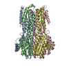 6iv1C 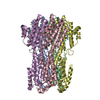 6iv2C  6iv4C  6ivjC  6ivkC  6ivlC 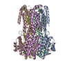 6ivmC 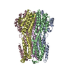 6ivnC 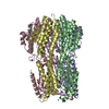 6ivoC  6ivpC 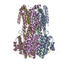 6ivqC 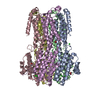 6ivrC  6ivwC  6jlfC C: citing same article ( |
|---|---|
| Similar structure data |


-Links
 PDBj
PDBj- Assembly
| Deposited unit | 
| ||||||||
|---|---|---|---|---|---|---|---|---|---|
| 1 |
| ||||||||
| Unit cell |
|
-Components
| #1: Protein | Mass: 33629.031 Da / Num. of mol.: 5 / Mutation: W252A Source method: isolated from a genetically manipulated source Source: (gene. exp.) Klebsiella pneumoniae IS53 (bacteria) / Production host: Escherichia coli (E. coli) / References: UniProt: W1ELP7#2: Chemical | ChemComp-ZN /   Mass: 65.409 Da / Num. of mol.: 15 / Source method: obtained synthetically / Formula: Zn Mass: 65.409 Da / Num. of mol.: 15 / Source method: obtained synthetically / Formula: Zn#3: Water | ChemComp-HOH / | Water Mass: 18.015 Da / Num. of mol.: 80 / Source method: isolated from a natural source / Formula: H2O Mass: 18.015 Da / Num. of mol.: 80 / Source method: isolated from a natural source / Formula: H2O |
|---|
-Experimental details
-Experiment
| Experiment | Method: X-RAY DIFFRACTION / Number of used crystals: 1 |
|---|
- Sample preparation
| Crystal | Density Matthews: 4.38 Å3/Da / Density % sol: 71.92 % |
|---|---|
| Crystal grow | Temperature: 293 K / Method: vapor diffusion, sitting drop Details: 0.05M zinc acetate, 6% v/v ethylene glycol, 0.1M sodium cacodylate, pH 6.0, 6.6% w/v PEG 8000 |
-Data collection
| Diffraction | Mean temperature: 100 K / Serial crystal experiment: N |
|---|---|
| Diffraction source | Source: SYNCHROTRON / Site: APS  / Beamline: 24-ID-E / Wavelength: 0.98 Å / Beamline: 24-ID-E / Wavelength: 0.98 Å |
| Detector | Type: DECTRIS EIGER X 16M / Detector: PIXEL / Date: Nov 20, 2018 |
| Radiation | Protocol: SINGLE WAVELENGTH / Monochromatic (M) / Laue (L): M / Scattering type: x-ray |
| Radiation wavelength | Wavelength: 0.98 Å / Relative weight: 1 |
| Reflection | Resolution: 2.52→66.11 Å / Num. obs: 99774 / % possible obs: 98.7 % / Redundancy: 3.4 % / Net I/σ(I): 12.75 |
| Reflection shell | Resolution: 2.52→2.61 Å |
- Processing
| Software |
| |||||||||||||||||||||||||||||||||||||||||||||||||||||||||||||||||||||||||||||||||||||||||||||||||||||||||
|---|---|---|---|---|---|---|---|---|---|---|---|---|---|---|---|---|---|---|---|---|---|---|---|---|---|---|---|---|---|---|---|---|---|---|---|---|---|---|---|---|---|---|---|---|---|---|---|---|---|---|---|---|---|---|---|---|---|---|---|---|---|---|---|---|---|---|---|---|---|---|---|---|---|---|---|---|---|---|---|---|---|---|---|---|---|---|---|---|---|---|---|---|---|---|---|---|---|---|---|---|---|---|---|---|---|---|
| Refinement | Resolution: 2.515→66.106 Å / SU ML: 0.38 / Cross valid method: FREE R-VALUE / σ(F): 1.35 / Phase error: 28.8 / Stereochemistry target values: ML
| |||||||||||||||||||||||||||||||||||||||||||||||||||||||||||||||||||||||||||||||||||||||||||||||||||||||||
| Solvent computation | Shrinkage radii: 0.9 Å / VDW probe radii: 1.11 Å / Solvent model: FLAT BULK SOLVENT MODEL | |||||||||||||||||||||||||||||||||||||||||||||||||||||||||||||||||||||||||||||||||||||||||||||||||||||||||
| Refinement step | Cycle: LAST / Resolution: 2.515→66.106 Å
| |||||||||||||||||||||||||||||||||||||||||||||||||||||||||||||||||||||||||||||||||||||||||||||||||||||||||
| Refine LS restraints |
| |||||||||||||||||||||||||||||||||||||||||||||||||||||||||||||||||||||||||||||||||||||||||||||||||||||||||
| LS refinement shell |
|
