 Movie
Movie Controller

[English] 日本語
 Yorodumi
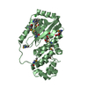
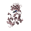
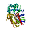
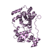
- PDB-6iuq: Crystal structure and expression patterns of prolyl 4-hydroxylase... -
+ Open data

- Basic information
| Entry | Database: PDB / ID: 6iuq | ||||||
|---|---|---|---|---|---|---|---|
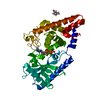
| Title | Crystal structure and expression patterns of prolyl 4-hydroxylases from Phytophthora capsici | ||||||
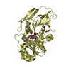
 Components Components | Prolyl 4-hydroxylase Procollagen-proline dioxygenase Procollagen-proline dioxygenase | ||||||
 Keywords Keywords | OXIDOREDUCTASE / hydroxylase / iron / DSBH core fold | ||||||
| Function / homology |  Function and homology informationL-ascorbic acid binding / dioxygenase activity / oxidoreductase activity, acting on paired donors, with incorporation or reduction of molecular oxygen / iron ion binding Function and homology informationL-ascorbic acid binding / dioxygenase activity / oxidoreductase activity, acting on paired donors, with incorporation or reduction of molecular oxygen / iron ion bindingSimilarity search - Function | ||||||
| Biological species |  Phytophthora capsici LT1534 (eukaryote) Phytophthora capsici LT1534 (eukaryote) | ||||||
| Method | X-RAY DIFFRACTION / SYNCHROTRON / SAD / Resolution: 2.348 Å | ||||||
 Authors Authors | Song, W.W. / Zhang, X.G. | ||||||
| Funding support |  China, 1items China, 1items
| ||||||
 Citation Citation | Journal: To Be Published Title: Crystal structure and expression patterns of prolyl 4-hydroxylases from Phytophthora capsici Authors: Song, W.W. / Zhang, X.G. | ||||||
| History |
|
- Structure visualization
| Structure viewer | Molecule: MolmilJmol/JSmol |
|---|
- Downloads & links
-Download
| PDBx/mmCIF format | 6iuq.cif.gz | 135.2 KB | Display | PDBx/mmCIF format |
|---|---|---|---|---|
| PDB format | pdb6iuq.ent.gz | 110.9 KB | Display | PDB format |
| PDBx/mmJSON format | 6iuq.json.gz | Tree view | PDBx/mmJSON format | |
| Others |  Other downloads Other downloads |
-Validation report
| Arichive directory | https://data.pdbj.org/pub/pdb/validation_reports/iu/6iuqftp://data.pdbj.org/pub/pdb/validation_reports/iu/6iuq | HTTPS FTP |
|---|
-Related structure data
| Similar structure data |
|---|


-Links
 PDBj
PDBj


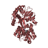
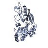
- Assembly
| Deposited unit | 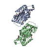
| ||||||||
|---|---|---|---|---|---|---|---|---|---|
| 1 | 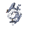
| ||||||||
| 2 | 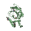
| ||||||||
| Unit cell |
|
-Components
| #1: Protein | Procollagen-proline dioxygenase Mass: 40481.461 Da / Num. of mol.: 2 Source method: isolated from a genetically manipulated source Details: each monomer consisits of an iron ion Source: (gene. exp.) Phytophthora capsici LT1534 (eukaryote)Production host:  Escherichia coli (E. coli) / References: UniProt: A0A4P1LYH8*PLUS Escherichia coli (E. coli) / References: UniProt: A0A4P1LYH8*PLUS#2: Chemical |   Mass: 55.845 Da / Num. of mol.: 2 / Source method: isolated from a natural source / Formula: Fe Mass: 55.845 Da / Num. of mol.: 2 / Source method: isolated from a natural source / Formula: Fe#3: Water | ChemComp-HOH / | Water Mass: 18.015 Da / Num. of mol.: 249 / Source method: isolated from a natural source / Formula: H2O Mass: 18.015 Da / Num. of mol.: 249 / Source method: isolated from a natural source / Formula: H2OSequence details | GenBank number of this polypeptide is MH251252. | |
|---|
-Experimental details
-Experiment
| Experiment | Method: X-RAY DIFFRACTION / Number of used crystals: 1 |
|---|
- Sample preparation
| Crystal | Density Matthews: 3.04 Å3/Da / Density % sol: 65.31 % |
|---|---|
| Crystal grow | Temperature: 283 K / Method: vapor diffusion, sitting drop Details: 0.1M sodium citrate tribasic dehydrate pH5.0, 18% (w/v) PEG20000 |
-Data collection
| Diffraction | Mean temperature: 100 K / Serial crystal experiment: N |
|---|---|
| Diffraction source | Source: SYNCHROTRON / Site: SSRF / Beamline: BL18U1 / Wavelength: 0.979 Å |
| Detector | Type: DECTRIS PILATUS3 6M / Detector: PIXEL / Date: Jan 22, 2018 |
| Radiation | Protocol: SINGLE WAVELENGTH / Monochromatic (M) / Laue (L): M / Scattering type: x-ray |
| Radiation wavelength | Wavelength: 0.979 Å / Relative weight: 1 |
| Reflection | Resolution: 2.348→50 Å / Num. obs: 38776 / % possible obs: 98 % / Redundancy: 5.3 % / Rpim(I) all: 0.029 / Net I/σ(I): 8.7 |
| Reflection shell | Resolution: 2.35→2.39 Å / Num. unique obs: 1882 / Rpim(I) all: 0.172 |
- Processing
| Software |
| ||||||||||||||||||||||||||||||||||||||||||||||||||||||||||||||||||||||||||||||||||||||||||||||||||
|---|---|---|---|---|---|---|---|---|---|---|---|---|---|---|---|---|---|---|---|---|---|---|---|---|---|---|---|---|---|---|---|---|---|---|---|---|---|---|---|---|---|---|---|---|---|---|---|---|---|---|---|---|---|---|---|---|---|---|---|---|---|---|---|---|---|---|---|---|---|---|---|---|---|---|---|---|---|---|---|---|---|---|---|---|---|---|---|---|---|---|---|---|---|---|---|---|---|---|---|
| Refinement | Method to determine structure: SAD / Resolution: 2.348→43.669 Å / SU ML: 0.22 / Cross valid method: NONE / σ(F): 1.98 / Phase error: 22.56
| ||||||||||||||||||||||||||||||||||||||||||||||||||||||||||||||||||||||||||||||||||||||||||||||||||
| Solvent computation | Shrinkage radii: 0.9 Å / VDW probe radii: 1.11 Å | ||||||||||||||||||||||||||||||||||||||||||||||||||||||||||||||||||||||||||||||||||||||||||||||||||
| Refinement step | Cycle: LAST / Resolution: 2.348→43.669 Å
| ||||||||||||||||||||||||||||||||||||||||||||||||||||||||||||||||||||||||||||||||||||||||||||||||||
| Refine LS restraints |
| ||||||||||||||||||||||||||||||||||||||||||||||||||||||||||||||||||||||||||||||||||||||||||||||||||
| LS refinement shell |
|
