 Movie
Movie Controller

[English] 日本語
 Yorodumi
- PDB-5kz7: Mark2 complex with 7-[(1S)-1-(4-fluorophenyl)ethyl]-5,5-dimethyl-... -
+ Open data

- Basic information
| Entry | Database: PDB / ID: 5kz7 | ||||||
|---|---|---|---|---|---|---|---|
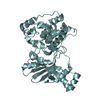
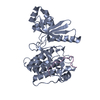
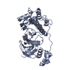
| Title | Mark2 complex with 7-[(1S)-1-(4-fluorophenyl)ethyl]-5,5-dimethyl-2-(3-pyridylamino)pyrrolo[2,3-d]pyrimidin-6-one | ||||||
 Components Components | Serine/threonine-protein kinase MARK2 | ||||||
 Keywords Keywords | Transferase/Transferase Inhibitor / Mark /  Serine/threonine-protein kinase / Transferase-Transferase Inhibitor complex Serine/threonine-protein kinase / Transferase-Transferase Inhibitor complex | ||||||
| Function / homology |  Function and homology information Function and homology informationestablishment or maintenance of cell polarity regulating cell shape / microtubule bundle / regulation of microtubule binding / mitochondrion localization / autophagy of mitochondrion / tau-protein kinase / establishment or maintenance of epithelial cell apical/basal polarity / regulation of axonogenesis / establishment of cell polarity / tau-protein kinase activity ...establishment or maintenance of cell polarity regulating cell shape / microtubule bundle / regulation of microtubule binding / mitochondrion localization / autophagy of mitochondrion / tau-protein kinase / establishment or maintenance of epithelial cell apical/basal polarity / regulation of axonogenesis / establishment of cell polarity / tau-protein kinase activity / axon development / activation of protein kinase activity / protein kinase activator activity / regulation of cytoskeleton organization / lateral plasma membrane / regulation of microtubule cytoskeleton organization / actin filament / peptidyl-threonine phosphorylation / neuron migration / tau protein binding / Wnt signaling pathway / microtubule cytoskeleton organization / positive regulation of neuron projection development / peptidyl-serine phosphorylation / protein autophosphorylation / non-specific serine/threonine protein kinase / intracellular signal transduction / cadherin binding / protein phosphorylation / protein serine kinase activity / protein serine/threonine kinase activity / lipid binding / dendrite / magnesium ion binding / mitochondrion / RNA binding / nucleoplasm / ATP binding / membrane / plasma membrane / cytoplasmSimilarity search - Function | ||||||
| Biological species |  Homo sapiens (human) Homo sapiens (human) | ||||||
| Method | X-RAY DIFFRACTION / FOURIER SYNTHESIS / Resolution: 3.2 Å | ||||||
 Authors Authors | Su, H.P. / Munshi, S.K. | ||||||
 Citation Citation | Journal: Bioorg. Med. Chem. Lett. / Year: 2017 Title: Structure guided design of a series of selective pyrrolopyrimidinone MARK inhibitors. Authors: Katz, J.D. / Haidle, A. / Childers, K.K. / Zabierek, A.A. / Jewell, J.P. / Hou, Y. / Altman, M.D. / Szewczak, A. / Chen, D. / Harsch, A. / Hayashi, M. / Warren, L. / Hutton, M. / Nuthall, H. ...Authors: Katz, J.D. / Haidle, A. / Childers, K.K. / Zabierek, A.A. / Jewell, J.P. / Hou, Y. / Altman, M.D. / Szewczak, A. / Chen, D. / Harsch, A. / Hayashi, M. / Warren, L. / Hutton, M. / Nuthall, H. / Su, H.P. / Munshi, S. / Stanton, M.G. / Davies, I.W. / Munoz, B. / Northrup, A. | ||||||
| History |
|
- Structure visualization
| Structure viewer | Molecule: MolmilJmol/JSmol |
|---|
- Downloads & links
-Download
| PDBx/mmCIF format | 5kz7.cif.gz | 130.8 KB | Display | PDBx/mmCIF format |
|---|---|---|---|---|
| PDB format | pdb5kz7.ent.gz | 100.9 KB | Display | PDB format |
| PDBx/mmJSON format | 5kz7.json.gz | Tree view | PDBx/mmJSON format | |
| Others |  Other downloads Other downloads |
-Validation report
| Arichive directory | https://data.pdbj.org/pub/pdb/validation_reports/kz/5kz7ftp://data.pdbj.org/pub/pdb/validation_reports/kz/5kz7 | HTTPS FTP |
|---|
-Related structure data
| Related structure data |  5kz8C  5eakS C: citing same article ( S: Starting model for refinement |
|---|---|
| Similar structure data |


-Links
 PDBj
PDBj- Assembly
| Deposited unit | 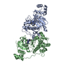
| ||||||||
|---|---|---|---|---|---|---|---|---|---|
| 1 | 
| ||||||||
| 2 | 
| ||||||||
| Unit cell |
|
-Components
| #1: Protein | Mass: 40056.090 Da / Num. of mol.: 2 / Fragment: UNP residues 6-331 Source method: isolated from a genetically manipulated source Source: (gene. exp.) Homo sapiens (human) / Gene: MARK2, EMK1 / Production host:  Escherichia coli (E. coli) Escherichia coli (E. coli)References: UniProt: Q7KZI7, non-specific serine/threonine protein kinase, tau-protein kinase#2: Chemical |   Mass: 377.415 Da / Num. of mol.: 2 / Source method: obtained synthetically / Formula: C21H20FN5O Mass: 377.415 Da / Num. of mol.: 2 / Source method: obtained synthetically / Formula: C21H20FN5O |
|---|
-Experimental details
-Experiment
| Experiment | Method: X-RAY DIFFRACTION / Number of used crystals: 1 |
|---|
- Sample preparation
| Crystal | Density Matthews: 2.53 Å3/Da / Density % sol: 51.46 % / Mosaicity: 0.809 ° |
|---|---|
| Crystal grow | Temperature: 298 K / Method: vapor diffusion, hanging drop / pH: 6.5 Details: 0.1M BIS-TRIS PH 6.5, 14% PEG3350, 200MM AMM.SULFATE |
-Data collection
| Diffraction | Mean temperature: 100 K | |||||||||||||||||||||||||||||||||||||||||||||||||||||||
|---|---|---|---|---|---|---|---|---|---|---|---|---|---|---|---|---|---|---|---|---|---|---|---|---|---|---|---|---|---|---|---|---|---|---|---|---|---|---|---|---|---|---|---|---|---|---|---|---|---|---|---|---|---|---|---|---|
| Diffraction source | Source: ROTATING ANODE / Type: RIGAKU MICROMAX-007 / Wavelength: 1.54 Å | |||||||||||||||||||||||||||||||||||||||||||||||||||||||
| Detector | Type: RIGAKU / Detector: IMAGE PLATE / Date: Oct 24, 2007 | |||||||||||||||||||||||||||||||||||||||||||||||||||||||
| Radiation | Protocol: SINGLE WAVELENGTH / Monochromatic (M) / Laue (L): M / Scattering type: x-ray | |||||||||||||||||||||||||||||||||||||||||||||||||||||||
| Radiation wavelength | Wavelength: 1.54 Å / Relative weight: 1 | |||||||||||||||||||||||||||||||||||||||||||||||||||||||
| Reflection | Resolution: 3.2→20 Å / Num. obs: 12561 / % possible obs: 69.6 % / Redundancy: 1.4 % / Biso Wilson estimate: 94.78 Å2 / Rmerge(I) obs: 0.131 / Χ2: 1.617 / Net I/av σ(I): 5.235 / Net I/σ(I): 5.6 / Num. measured all: 11068 | |||||||||||||||||||||||||||||||||||||||||||||||||||||||
| Reflection shell |
|
- Processing
| Software |
| ||||||||||||||||||||||||||||||||||||||||||||||||||||||||||||||||||||||||||||||||||||||||||||||||||||||||||||
|---|---|---|---|---|---|---|---|---|---|---|---|---|---|---|---|---|---|---|---|---|---|---|---|---|---|---|---|---|---|---|---|---|---|---|---|---|---|---|---|---|---|---|---|---|---|---|---|---|---|---|---|---|---|---|---|---|---|---|---|---|---|---|---|---|---|---|---|---|---|---|---|---|---|---|---|---|---|---|---|---|---|---|---|---|---|---|---|---|---|---|---|---|---|---|---|---|---|---|---|---|---|---|---|---|---|---|---|---|---|
| Refinement | Method to determine structure: FOURIER SYNTHESIS Starting model: 5EAK Resolution: 3.2→19.53 Å / Cor.coef. Fo:Fc: 0.8876 / Cor.coef. Fo:Fc free: 0.8031 / Cross valid method: THROUGHOUT / σ(F): 0 / SU Rfree Blow DPI: 0.564
| ||||||||||||||||||||||||||||||||||||||||||||||||||||||||||||||||||||||||||||||||||||||||||||||||||||||||||||
| Displacement parameters | Biso max: 144.25 Å2 / Biso mean: 72.41 Å2 / Biso min: 19.35 Å2
| ||||||||||||||||||||||||||||||||||||||||||||||||||||||||||||||||||||||||||||||||||||||||||||||||||||||||||||
| Refine analyze | Luzzati coordinate error obs: 0.615 Å | ||||||||||||||||||||||||||||||||||||||||||||||||||||||||||||||||||||||||||||||||||||||||||||||||||||||||||||
| Refinement step | Cycle: final / Resolution: 3.2→19.53 Å
| ||||||||||||||||||||||||||||||||||||||||||||||||||||||||||||||||||||||||||||||||||||||||||||||||||||||||||||
| Refine LS restraints |
| ||||||||||||||||||||||||||||||||||||||||||||||||||||||||||||||||||||||||||||||||||||||||||||||||||||||||||||
| LS refinement shell | Resolution: 3.2→3.5 Å / Total num. of bins used: 6
|
