 Movie
Movie Controller

[English] 日本語
 Yorodumi
- PDB-6i4u: Crystal structure of the disease-causing G426E mutant of the huma... -
+ Open data

- Basic information
| Entry | Database: PDB / ID: 6i4u | |||||||||||||||||||||||||||||||||
|---|---|---|---|---|---|---|---|---|---|---|---|---|---|---|---|---|---|---|---|---|---|---|---|---|---|---|---|---|---|---|---|---|---|---|
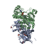
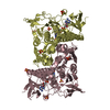
| Title | Crystal structure of the disease-causing G426E mutant of the human dihydrolipoamide dehydrogenase | |||||||||||||||||||||||||||||||||
 Components Components | Dihydrolipoyl dehydrogenase, mitochondrial Dihydrolipoamide dehydrogenase Dihydrolipoamide dehydrogenase | |||||||||||||||||||||||||||||||||
 Keywords Keywords | OXIDOREDUCTASE / Lipoamide dehydrogenase / Pathogenic mutation / E3 deficiency / Alpha-ketoglutarate dehydrogenase complex / 2-oxoglutarate dehydrogenase complex / Pyruvate dehydrogenase complex | |||||||||||||||||||||||||||||||||
| Function / homology |  Function and homology informationacetyltransferase complex / acrosomal matrix / Glycine degradation / : / dihydrolipoyl dehydrogenase / dihydrolipoyl dehydrogenase activity / oxoglutarate dehydrogenase complex / acetyl-CoA biosynthetic process from pyruvate / pyruvate dehydrogenase complex / : ...acetyltransferase complex / acrosomal matrix / Glycine degradation / : / dihydrolipoyl dehydrogenase / dihydrolipoyl dehydrogenase activity / oxoglutarate dehydrogenase complex / acetyl-CoA biosynthetic process from pyruvate / pyruvate dehydrogenase complex / : / Lysine catabolism / branched-chain amino acid catabolic process / Citric acid cycle (TCA cycle) / Branched-chain amino acid catabolism / Pyruvate metabolism / Glyoxylate metabolism and glycine degradation / Regulation of pyruvate dehydrogenase (PDH) complex / motile cilium / sperm capacitation / Signaling by Retinoic Acid / mitochondrial electron transport, NADH to ubiquinone / Mitochondrial protein degradation / gastrulation / regulation of membrane potential / flavin adenine dinucleotide binding / mitochondrial matrix / mitochondrion / proteolysis / nucleus Function and homology informationacetyltransferase complex / acrosomal matrix / Glycine degradation / : / dihydrolipoyl dehydrogenase / dihydrolipoyl dehydrogenase activity / oxoglutarate dehydrogenase complex / acetyl-CoA biosynthetic process from pyruvate / pyruvate dehydrogenase complex / : ...acetyltransferase complex / acrosomal matrix / Glycine degradation / : / dihydrolipoyl dehydrogenase / dihydrolipoyl dehydrogenase activity / oxoglutarate dehydrogenase complex / acetyl-CoA biosynthetic process from pyruvate / pyruvate dehydrogenase complex / : / Lysine catabolism / branched-chain amino acid catabolic process / Citric acid cycle (TCA cycle) / Branched-chain amino acid catabolism / Pyruvate metabolism / Glyoxylate metabolism and glycine degradation / Regulation of pyruvate dehydrogenase (PDH) complex / motile cilium / sperm capacitation / Signaling by Retinoic Acid / mitochondrial electron transport, NADH to ubiquinone / Mitochondrial protein degradation / gastrulation / regulation of membrane potential / flavin adenine dinucleotide binding / mitochondrial matrix / mitochondrion / proteolysis / nucleusSimilarity search - Function | |||||||||||||||||||||||||||||||||
| Biological species |  Homo sapiens (human) Homo sapiens (human) | |||||||||||||||||||||||||||||||||
| Method | X-RAY DIFFRACTION / SYNCHROTRON / MOLECULAR REPLACEMENT / molecular replacement / Resolution: 1.84 Å | |||||||||||||||||||||||||||||||||
 Authors Authors | Szabo, E. / Wilk, P. / Hubert, A. / Torocsik, B. / Weiss, M.S. / Adam-Vizi, V. / Ambrus, A. | |||||||||||||||||||||||||||||||||
| Funding support |  Hungary, Hungary,  United States, United States,  Germany, 10items Germany, 10items
| |||||||||||||||||||||||||||||||||
 Citation Citation | Journal: Hum.Mol.Genet. / Year: 2019 Title: Underlying molecular alterations in human dihydrolipoamide dehydrogenase deficiency revealed by structural analyses of disease-causing enzyme variants. Authors: Szabo, E. / Wilk, P. / Nagy, B. / Zambo, Z. / Bui, D. / Weichsel, A. / Arjunan, P. / Torocsik, B. / Hubert, A. / Furey, W. / Montfort, W.R. / Jordan, F. / Weiss, M.S. / Adam-Vizi, V. / Ambrus, A. | |||||||||||||||||||||||||||||||||
| History |
|
- Structure visualization
| Structure viewer | Molecule: MolmilJmol/JSmol |
|---|
- Downloads & links
-Download
| PDBx/mmCIF format | 6i4u.cif.gz | 542.1 KB | Display | PDBx/mmCIF format |
|---|---|---|---|---|
| PDB format | pdb6i4u.ent.gz | 453.7 KB | Display | PDB format |
| PDBx/mmJSON format | 6i4u.json.gz | Tree view | PDBx/mmJSON format | |
| Others |  Other downloads Other downloads |
-Validation report
| Arichive directory | https://data.pdbj.org/pub/pdb/validation_reports/i4/6i4uftp://data.pdbj.org/pub/pdb/validation_reports/i4/6i4u | HTTPS FTP |
|---|
-Related structure data
| Related structure data |  6i4pC  6i4qC  6i4rC 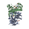 6i4sC 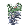 6i4tC  6i4zC  1zmdS S: Starting model for refinement C: citing same article ( |
|---|---|
| Similar structure data |


-Links
 PDBj
PDBj


- Assembly
| Deposited unit | 
| ||||||||
|---|---|---|---|---|---|---|---|---|---|
| 1 |
| ||||||||
| Unit cell |
|
-Components
| #1: Protein | Dihydrolipoamide dehydrogenase / Dihydrolipoamide dehydrogenase / Glycine cleavage system L protein Mass: 52709.234 Da / Num. of mol.: 2 / Mutation: G426E Source method: isolated from a genetically manipulated source Details: Sequence of the Strep-tag with linker amino acids: MASWSHPQFEKGALEVLFQGPG Source: (gene. exp.) Homo sapiens (human) / Gene: DLD, GCSL, LAD, PHE3 / Plasmid: pET52b+ / Production host:  Escherichia coli BL21(DE3) (bacteria) / References: UniProt: P09622, dihydrolipoyl dehydrogenase Escherichia coli BL21(DE3) (bacteria) / References: UniProt: P09622, dihydrolipoyl dehydrogenase#2: Chemical | Flavin adenine dinucleotide  Mass: 785.550 Da / Num. of mol.: 2 / Source method: obtained synthetically / Formula: C27H33N9O15P2 / Comment: FAD*YM Mass: 785.550 Da / Num. of mol.: 2 / Source method: obtained synthetically / Formula: C27H33N9O15P2 / Comment: FAD*YM#3: Chemical | ChemComp-SO4 / Sulfate  Mass: 96.063 Da / Num. of mol.: 9 / Source method: obtained synthetically / Formula: SO4 Mass: 96.063 Da / Num. of mol.: 9 / Source method: obtained synthetically / Formula: SO4#4: Chemical | ChemComp-1PE / | Polyethylene glycol  Mass: 238.278 Da / Num. of mol.: 1 / Source method: obtained synthetically / Formula: C10H22O6 / Comment: precipitant*YM Mass: 238.278 Da / Num. of mol.: 1 / Source method: obtained synthetically / Formula: C10H22O6 / Comment: precipitant*YM#5: Water | ChemComp-HOH / | Water Mass: 18.015 Da / Num. of mol.: 347 / Source method: isolated from a natural source / Formula: H2O Mass: 18.015 Da / Num. of mol.: 347 / Source method: isolated from a natural source / Formula: H2O |
|---|
-Experimental details
-Experiment
| Experiment | Method: X-RAY DIFFRACTION / Number of used crystals: 1 |
|---|
- Sample preparation
| Crystal | Density Matthews: 3 Å3/Da / Density % sol: 58.99 % |
|---|---|
| Crystal grow | Temperature: 293 K / Method: vapor diffusion, sitting drop / pH: 7.5 Details: 2 M ammonium sulfate, 2(v/v)% PEG 400, 0.1 M Hepes (pH 7.5) |
-Data collection
| Diffraction | Mean temperature: 100 K / Serial crystal experiment: N | ||||||||||||||||||||||||||||||||||||||||||||||||||||||||||||||||||||||||||||||||
|---|---|---|---|---|---|---|---|---|---|---|---|---|---|---|---|---|---|---|---|---|---|---|---|---|---|---|---|---|---|---|---|---|---|---|---|---|---|---|---|---|---|---|---|---|---|---|---|---|---|---|---|---|---|---|---|---|---|---|---|---|---|---|---|---|---|---|---|---|---|---|---|---|---|---|---|---|---|---|---|---|---|
| Diffraction source | Source: SYNCHROTRON / Site: BESSY / Beamline: 14.1 / Wavelength: 0.9184 Å | ||||||||||||||||||||||||||||||||||||||||||||||||||||||||||||||||||||||||||||||||
| Detector | Type: DECTRIS PILATUS 6M / Detector: PIXEL / Date: Sep 24, 2016 | ||||||||||||||||||||||||||||||||||||||||||||||||||||||||||||||||||||||||||||||||
| Radiation | Protocol: SINGLE WAVELENGTH / Monochromatic (M) / Laue (L): M / Scattering type: x-ray | ||||||||||||||||||||||||||||||||||||||||||||||||||||||||||||||||||||||||||||||||
| Radiation wavelength | Wavelength: 0.9184 Å / Relative weight: 1 | ||||||||||||||||||||||||||||||||||||||||||||||||||||||||||||||||||||||||||||||||
| Reflection | Resolution: 1.84→42.559 Å / Num. obs: 106853 / % possible obs: 99.2 % / Redundancy: 6.658 % / Biso Wilson estimate: 28.8 Å2 / CC1/2: 0.999 / Rmerge(I) obs: 0.098 / Rrim(I) all: 0.106 / Χ2: 1.13 / Net I/σ(I): 13.27 | ||||||||||||||||||||||||||||||||||||||||||||||||||||||||||||||||||||||||||||||||
| Reflection shell | Diffraction-ID: 1
|
-Phasing
| Phasing | Method: molecular replacement | ||||||
|---|---|---|---|---|---|---|---|
| Phasing MR | R rigid body: 0.604
|
- Processing
| Software |
| ||||||||||||||||||||||||||||||||||||||||||||||||||||||||||||||||||||||||||||||||||||||||||||||||||||||||||||||||||||||||||||||||||||||||||||||||||||||||||||||||||||||||||||||||||||||||||||||||||||||||||||||||||||||||||||||||||||||||||||||||||||||||||
|---|---|---|---|---|---|---|---|---|---|---|---|---|---|---|---|---|---|---|---|---|---|---|---|---|---|---|---|---|---|---|---|---|---|---|---|---|---|---|---|---|---|---|---|---|---|---|---|---|---|---|---|---|---|---|---|---|---|---|---|---|---|---|---|---|---|---|---|---|---|---|---|---|---|---|---|---|---|---|---|---|---|---|---|---|---|---|---|---|---|---|---|---|---|---|---|---|---|---|---|---|---|---|---|---|---|---|---|---|---|---|---|---|---|---|---|---|---|---|---|---|---|---|---|---|---|---|---|---|---|---|---|---|---|---|---|---|---|---|---|---|---|---|---|---|---|---|---|---|---|---|---|---|---|---|---|---|---|---|---|---|---|---|---|---|---|---|---|---|---|---|---|---|---|---|---|---|---|---|---|---|---|---|---|---|---|---|---|---|---|---|---|---|---|---|---|---|---|---|---|---|---|---|---|---|---|---|---|---|---|---|---|---|---|---|---|---|---|---|---|---|---|---|---|---|---|---|---|---|---|---|---|---|---|---|---|---|---|---|---|---|---|---|---|---|---|---|---|---|---|---|---|
| Refinement | Method to determine structure: MOLECULAR REPLACEMENT Starting model: 1ZMD Resolution: 1.84→42.559 Å / SU ML: 0.21 / Cross valid method: THROUGHOUT / σ(F): 1.36 / Phase error: 24.97 Details: TLS groups, NCS torsion-angle restraints, automatic occupancy refinement and real-space refinement were involved, hydrogen atoms were added to the final model during refinement
| ||||||||||||||||||||||||||||||||||||||||||||||||||||||||||||||||||||||||||||||||||||||||||||||||||||||||||||||||||||||||||||||||||||||||||||||||||||||||||||||||||||||||||||||||||||||||||||||||||||||||||||||||||||||||||||||||||||||||||||||||||||||||||
| Solvent computation | Shrinkage radii: 0.9 Å / VDW probe radii: 1.11 Å | ||||||||||||||||||||||||||||||||||||||||||||||||||||||||||||||||||||||||||||||||||||||||||||||||||||||||||||||||||||||||||||||||||||||||||||||||||||||||||||||||||||||||||||||||||||||||||||||||||||||||||||||||||||||||||||||||||||||||||||||||||||||||||
| Refinement step | Cycle: LAST / Resolution: 1.84→42.559 Å
| ||||||||||||||||||||||||||||||||||||||||||||||||||||||||||||||||||||||||||||||||||||||||||||||||||||||||||||||||||||||||||||||||||||||||||||||||||||||||||||||||||||||||||||||||||||||||||||||||||||||||||||||||||||||||||||||||||||||||||||||||||||||||||
| Refine LS restraints |
| ||||||||||||||||||||||||||||||||||||||||||||||||||||||||||||||||||||||||||||||||||||||||||||||||||||||||||||||||||||||||||||||||||||||||||||||||||||||||||||||||||||||||||||||||||||||||||||||||||||||||||||||||||||||||||||||||||||||||||||||||||||||||||
| LS refinement shell |
| ||||||||||||||||||||||||||||||||||||||||||||||||||||||||||||||||||||||||||||||||||||||||||||||||||||||||||||||||||||||||||||||||||||||||||||||||||||||||||||||||||||||||||||||||||||||||||||||||||||||||||||||||||||||||||||||||||||||||||||||||||||||||||
| Refinement TLS params. | Method: refined / Refine-ID: X-RAY DIFFRACTION
| ||||||||||||||||||||||||||||||||||||||||||||||||||||||||||||||||||||||||||||||||||||||||||||||||||||||||||||||||||||||||||||||||||||||||||||||||||||||||||||||||||||||||||||||||||||||||||||||||||||||||||||||||||||||||||||||||||||||||||||||||||||||||||
| Refinement TLS group |
|
