 Movie
Movie Controller

[English] 日本語
 Yorodumi
- PDB-6i0r: Structure of quinolinate synthase in complex with 5-mercaptopyrid... -
+ Open data

- Basic information
| Entry | Database: PDB / ID: 6i0r | ||||||
|---|---|---|---|---|---|---|---|
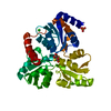
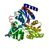
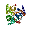
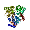
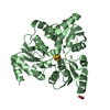
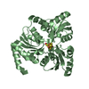
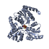
| Title | Structure of quinolinate synthase in complex with 5-mercaptopyridine-2,3-dicarboxylic acid | ||||||
 Components Components | Quinolinate synthase A | ||||||
 Keywords Keywords |  TRANSFERASE / NAD BIOSYNTHESIS / IRON SULFUR CLUSTER TRANSFERASE / NAD BIOSYNTHESIS / IRON SULFUR CLUSTER | ||||||
| Function / homology |  Function and homology information Function and homology information'de novo' NAD biosynthetic process from aspartate / quinolinate synthase / quinolinate synthetase A activity / 4 iron, 4 sulfur cluster binding / metal ion binding / cytosolSimilarity search - Function | ||||||
| Biological species |   Thermotoga maritima MSB8 (bacteria) Thermotoga maritima MSB8 (bacteria) | ||||||
| Method | X-RAY DIFFRACTION / SYNCHROTRON / MOLECULAR REPLACEMENT / molecular replacement / Resolution: 2.1 Å | ||||||
 Authors Authors | Volbeda, A. / Fontecilla-Camps, J.C. | ||||||
| Funding support |  France, 1items France, 1items
| ||||||
 Citation Citation | Journal: Chem.Commun.(Camb.) / Year: 2019 Title: Design of specific inhibitors of quinolinate synthase based on [4Fe-4S] cluster coordination. Authors: Saez Cabodevilla, J. / Volbeda, A. / Hamelin, O. / Latour, J.M. / Gigarel, O. / Clemancey, M. / Darnault, C. / Reichmann, D. / Amara, P. / Fontecilla-Camps, J.C. / Ollagnier de Choudens, S. | ||||||
| History |
|
- Structure visualization
| Structure viewer | Molecule: MolmilJmol/JSmol |
|---|
- Downloads & links
-Download
| PDBx/mmCIF format | 6i0r.cif.gz | 137 KB | Display | PDBx/mmCIF format |
|---|---|---|---|---|
| PDB format | pdb6i0r.ent.gz | 106 KB | Display | PDB format |
| PDBx/mmJSON format | 6i0r.json.gz | Tree view | PDBx/mmJSON format | |
| Others |  Other downloads Other downloads |
-Validation report
| Arichive directory | https://data.pdbj.org/pub/pdb/validation_reports/i0/6i0rftp://data.pdbj.org/pub/pdb/validation_reports/i0/6i0r | HTTPS FTP |
|---|
-Related structure data
| Related structure data |  6i0kC  6i0pC  6f48S C: citing same article ( S: Starting model for refinement |
|---|---|
| Similar structure data |


-Links
 PDBj
PDBj- Assembly
| Deposited unit | 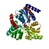
| ||||||||
|---|---|---|---|---|---|---|---|---|---|
| 1 |
| ||||||||
| Unit cell |
|
-Components
| #1: Protein | Mass: 34640.598 Da / Num. of mol.: 1 / Mutation: Y21F, K219R Source method: isolated from a genetically manipulated source Source: (gene. exp.) Thermotoga maritima MSB8 (bacteria) / Gene: nadA, TM_1644 / Plasmid: PT7 / Production host: Escherichia coli BL21(DE3) (bacteria) / References: UniProt: Q9X1X7, quinolinate synthase | ||||||
|---|---|---|---|---|---|---|---|
| #2: Chemical | ChemComp-FE / Iron  Mass: 55.845 Da / Num. of mol.: 4 / Source method: obtained synthetically / Formula: Fe Mass: 55.845 Da / Num. of mol.: 4 / Source method: obtained synthetically / Formula: Fe#3: Chemical | ChemComp-H2S / Hydrogen sulfide  Mass: 34.081 Da / Num. of mol.: 4 / Source method: obtained synthetically / Formula: H2S Mass: 34.081 Da / Num. of mol.: 4 / Source method: obtained synthetically / Formula: H2S#4: Chemical | ChemComp-QAT / |   Mass: 199.184 Da / Num. of mol.: 1 / Source method: obtained synthetically / Formula: C7H5NO4S Mass: 199.184 Da / Num. of mol.: 1 / Source method: obtained synthetically / Formula: C7H5NO4S#5: Water | ChemComp-HOH / | Water Mass: 18.015 Da / Num. of mol.: 110 / Source method: isolated from a natural source / Formula: H2O Mass: 18.015 Da / Num. of mol.: 110 / Source method: isolated from a natural source / Formula: H2O |
-Experimental details
-Experiment
| Experiment | Method: X-RAY DIFFRACTION / Number of used crystals: 2 |
|---|
- Sample preparation
| Crystal | Density Matthews: 2.24 Å3/Da / Density % sol: 45.19 % |
|---|---|
| Crystal grow | Temperature: 298 K / Method: vapor diffusion, hanging drop / pH: 6.7 / Details: PEG33500, dioxane, Na2HPO4, MES, anaerobic |
-Data collection
| Diffraction | Mean temperature: 100 K / Serial crystal experiment: N | ||||||||||||||||||||||||
|---|---|---|---|---|---|---|---|---|---|---|---|---|---|---|---|---|---|---|---|---|---|---|---|---|---|
| Diffraction source | Source: SYNCHROTRON / Site: ESRF / Beamline: ID30B / Wavelength: 1.00394 Å | ||||||||||||||||||||||||
| Detector | Type: DECTRIS PILATUS3 S 6M / Detector: PIXEL / Date: Jun 28, 2017 | ||||||||||||||||||||||||
| Radiation | Protocol: SINGLE WAVELENGTH / Monochromatic (M) / Laue (L): M / Scattering type: x-ray | ||||||||||||||||||||||||
| Radiation wavelength | Wavelength: 1.00394 Å / Relative weight: 1 | ||||||||||||||||||||||||
| Reflection | Resolution: 2.1→37.16 Å / Num. obs: 17011 / % possible obs: 95.3 % / Redundancy: 3 % / CC1/2: 0.982 / Rmerge(I) obs: 0.143 / Rpim(I) all: 0.091 / Rrim(I) all: 0.171 / Net I/σ(I): 8.5 / Num. measured all: 50636 | ||||||||||||||||||||||||
| Reflection shell | Diffraction-ID: 1
|
-Phasing
| Phasing | Method: molecular replacement |
|---|
- Processing
| Software |
| ||||||||||||||||||||||||||||||||||||||||||||||||||||||||||||||||||||||||||||||||||||||||||||||||||||
|---|---|---|---|---|---|---|---|---|---|---|---|---|---|---|---|---|---|---|---|---|---|---|---|---|---|---|---|---|---|---|---|---|---|---|---|---|---|---|---|---|---|---|---|---|---|---|---|---|---|---|---|---|---|---|---|---|---|---|---|---|---|---|---|---|---|---|---|---|---|---|---|---|---|---|---|---|---|---|---|---|---|---|---|---|---|---|---|---|---|---|---|---|---|---|---|---|---|---|---|---|---|
| Refinement | Method to determine structure: MOLECULAR REPLACEMENT Starting model: 6F48 Resolution: 2.1→37.16 Å / Cor.coef. Fo:Fc: 0.958 / Cor.coef. Fo:Fc free: 0.932 / SU B: 15.437 / SU ML: 0.197 / Cross valid method: THROUGHOUT / σ(F): 0 / ESU R: 0.28 / ESU R Free: 0.206 Details: U VALUES : WITH TLS ADDED HYDROGENS HAVE BEEN ADDED IN THE RIDING POSITIONS
| ||||||||||||||||||||||||||||||||||||||||||||||||||||||||||||||||||||||||||||||||||||||||||||||||||||
| Solvent computation | Ion probe radii: 0.8 Å / Shrinkage radii: 0.8 Å / VDW probe radii: 1.2 Å | ||||||||||||||||||||||||||||||||||||||||||||||||||||||||||||||||||||||||||||||||||||||||||||||||||||
| Displacement parameters | Biso max: 124.58 Å2 / Biso mean: 43.712 Å2 / Biso min: 21.98 Å2
| ||||||||||||||||||||||||||||||||||||||||||||||||||||||||||||||||||||||||||||||||||||||||||||||||||||
| Refinement step | Cycle: final / Resolution: 2.1→37.16 Å
| ||||||||||||||||||||||||||||||||||||||||||||||||||||||||||||||||||||||||||||||||||||||||||||||||||||
| Refine LS restraints |
| ||||||||||||||||||||||||||||||||||||||||||||||||||||||||||||||||||||||||||||||||||||||||||||||||||||
| LS refinement shell | Resolution: 2.1→2.154 Å / Rfactor Rfree error: 0 / Total num. of bins used: 20
| ||||||||||||||||||||||||||||||||||||||||||||||||||||||||||||||||||||||||||||||||||||||||||||||||||||
| Refinement TLS params. | Method: refined / Refine-ID: X-RAY DIFFRACTION
| ||||||||||||||||||||||||||||||||||||||||||||||||||||||||||||||||||||||||||||||||||||||||||||||||||||
| Refinement TLS group |
|
