 Movie
Movie Controller

[English] 日本語
 Yorodumi
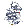
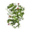
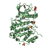
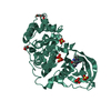
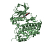
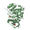
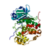
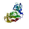
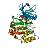
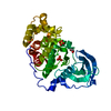
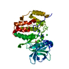
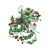
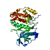
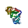
- PDB-6hme: LOW-SALT STRUCTURE OF PROTEIN KINASE CK2 CATALYTIC SUBUNIT (ISOFO... -
+ Open data

- Basic information
| Entry | Database: PDB / ID: 6hme | ||||||
|---|---|---|---|---|---|---|---|
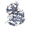
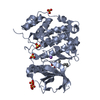
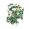
| Title | LOW-SALT STRUCTURE OF PROTEIN KINASE CK2 CATALYTIC SUBUNIT (ISOFORM CK2ALPHA; CSNK2A1 gene product) IN COMPLEX WITH THE INDENOINDOLE-TYPE INHIBITOR THN27 | ||||||
 Components Components | Casein kinase II subunit alpha Casein kinase 2 Casein kinase 2 | ||||||
 Keywords Keywords | TRANSFERASE / protein kinase CK2 / casein kinase 2 / catalytic subunit CK2alpha / CSNK2A1 / indenoindole-type inhibitor | ||||||
| Function / homology |  Function and homology information Function and homology informationregulation of chromosome separation / positive regulation of aggrephagy / WNT mediated activation of DVL / Condensation of Prometaphase Chromosomes / protein kinase CK2 complex / symbiont-mediated disruption of host cell PML body / Maturation of hRSV A proteins / Receptor Mediated Mitophagy / Sin3-type complex / Synthesis of PC ...regulation of chromosome separation / positive regulation of aggrephagy / WNT mediated activation of DVL / Condensation of Prometaphase Chromosomes / protein kinase CK2 complex / symbiont-mediated disruption of host cell PML body / Maturation of hRSV A proteins / Receptor Mediated Mitophagy / Sin3-type complex / Synthesis of PC / RUNX1 interacts with co-factors whose precise effect on RUNX1 targets is not known / negative regulation of apoptotic signaling pathway / positive regulation of Wnt signaling pathway / negative regulation of double-strand break repair via homologous recombination / chaperone-mediated protein folding / negative regulation of ubiquitin-dependent protein catabolic process / Signal transduction by L1 / peptidyl-threonine phosphorylation / Hsp90 protein binding / negative regulation of cysteine-type endopeptidase activity involved in apoptotic process / PML body / Wnt signaling pathway / Regulation of PTEN stability and activity / Cooperation of PDCL (PhLP1) and TRiC/CCT in G-protein beta folding / positive regulation of protein catabolic process / KEAP1-NFE2L2 pathway / double-strand break repair / rhythmic process / kinase activity / positive regulation of cell growth / peptidyl-serine phosphorylation / Regulation of TP53 Activity through Phosphorylation / negative regulation of translation / protein stabilization / non-specific serine/threonine protein kinase / regulation of cell cycle / cell cycle / protein phosphorylation / protein serine kinase activity / protein serine/threonine kinase activity / apoptotic process / DNA damage response / positive regulation of cell population proliferation / signal transduction / nucleoplasm / ATP binding / identical protein binding / nucleus / plasma membrane / cytosolSimilarity search - Function | ||||||
| Biological species |  Homo sapiens (human) Homo sapiens (human) | ||||||
| Method | X-RAY DIFFRACTION / SYNCHROTRON / MOLECULAR REPLACEMENT / Resolution: 1.85 Å | ||||||
 Authors Authors | Niefind, K. / Lindenblatt, D. / Jose, J. / Le Borgne, M. | ||||||
| Funding support |  Germany, 1items Germany, 1items
| ||||||
 Citation Citation | Journal: Acs Omega / Year: 2019 Title: Diacritic Binding of an Indenoindole Inhibitor by CK2 alpha Paralogs Explored by a Reliable Path to Atomic Resolution CK2 alpha ' Structures. Authors: Lindenblatt, D. / Nickelsen, A. / Applegate, V.M. / Hochscherf, J. / Witulski, B. / Bouaziz, Z. / Marminon, C. / Bretner, M. / Le Borgne, M. / Jose, J. / Niefind, K. #1: Journal: Pharmaceuticals (Basel) / Year: 2017Title: Unexpected Binding Mode of a Potent Indeno[1,2-b]indole-Type Inhibitor of Protein Kinase CK2 Revealed by Complex Structures with the Catalytic Subunit CK2alpha and Its Paralog CK2alpha' Authors: Hochscherf, J. / Lindenblatt, D. / Witulski, B. / Birus, R. / Aichele, D. / Marminon, C. / Bouaziz, Z. / Le Borgne, M. / Jose, J. / Niefind, K. #2: Journal: J. Mol. Biol. / Year: 2003Title: Crystal structure of a C-terminal deletion mutant of human protein kinase CK2 catalytic subunit. Authors: Ermakova, I. / Boldyreff, B. / Issinger, O.G. / Niefind, K. | ||||||
| History |
|
- Structure visualization
| Structure viewer | Molecule: MolmilJmol/JSmol |
|---|
- Downloads & links
-Download
| PDBx/mmCIF format | 6hme.cif.gz | 306.5 KB | Display | PDBx/mmCIF format |
|---|---|---|---|---|
| PDB format | pdb6hme.ent.gz | 249.5 KB | Display | PDB format |
| PDBx/mmJSON format | 6hme.json.gz | Tree view | PDBx/mmJSON format | |
| Others |  Other downloads Other downloads |
-Validation report
| Arichive directory | https://data.pdbj.org/pub/pdb/validation_reports/hm/6hmeftp://data.pdbj.org/pub/pdb/validation_reports/hm/6hme | HTTPS FTP |
|---|
-Related structure data
| Related structure data |  6hbnC  6hmbC  6hmcC  6hmdC 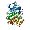 6hmqC  5oniS S: Starting model for refinement C: citing same article ( |
|---|---|
| Similar structure data |


-Links
 PDBj
PDBj


- Assembly
| Deposited unit | 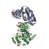
| ||||||||
|---|---|---|---|---|---|---|---|---|---|
| 1 | 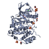
| ||||||||
| 2 | 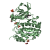
| ||||||||
| Unit cell |
|
-Components
-Protein , 1 types, 2 molecules AB
| #1: Protein | Casein kinase 2 / CK II alpha Mass: 41685.426 Da / Num. of mol.: 2 Source method: isolated from a genetically manipulated source Source: (gene. exp.) Homo sapiens (human) / Gene: CSNK2A1, CK2A1 / Production host:  Escherichia coli (E. coli) Escherichia coli (E. coli)References: UniProt: P68400, non-specific serine/threonine protein kinase |
|---|
-Non-polymers , 6 types, 409 molecules 


| #2: Chemical | ChemComp-EDO / Ethylene glycol Mass: 62.068 Da / Num. of mol.: 6 / Source method: obtained synthetically / Formula: C2H6O2 Mass: 62.068 Da / Num. of mol.: 6 / Source method: obtained synthetically / Formula: C2H6O2#3: Chemical | ChemComp-CL / | Chloride Mass: 35.453 Da / Num. of mol.: 1 / Source method: obtained synthetically / Formula: Cl Mass: 35.453 Da / Num. of mol.: 1 / Source method: obtained synthetically / Formula: Cl#4: Chemical | ChemComp-SO4 / Sulfate Mass: 96.063 Da / Num. of mol.: 10 / Source method: isolated from a natural source / Formula: SO4 Mass: 96.063 Da / Num. of mol.: 10 / Source method: isolated from a natural source / Formula: SO4#5: Chemical |  Mass: 335.396 Da / Num. of mol.: 2 / Source method: isolated from a natural source / Formula: C21H21NO3 Mass: 335.396 Da / Num. of mol.: 2 / Source method: isolated from a natural source / Formula: C21H21NO3#6: Chemical | ChemComp-NA / |  Mass: 22.990 Da / Num. of mol.: 1 / Source method: isolated from a natural source / Formula: Na Mass: 22.990 Da / Num. of mol.: 1 / Source method: isolated from a natural source / Formula: Na#7: Water | ChemComp-HOH / | WaterMass: 18.015 Da / Num. of mol.: 389 / Source method: isolated from a natural source / Formula: H2O |
|---|
-Experimental details
-Experiment
| Experiment | Method: X-RAY DIFFRACTION / Number of used crystals: 1 |
|---|
- Sample preparation
| Crystal | Density Matthews: 3.11 Å3/Da / Density % sol: 60.39 % |
|---|---|
| Crystal grow | Temperature: 293.15 K / Method: vapor diffusion, sitting drop / pH: 6.5 Details: 180 MICROLITERS OF ENZYME SOLUTION (6 MG/ML CK2ALPHA, 0.025 M TRIS/HCL, PH 8.5, 0.5 M NACL) WERE MIXED WITH 20 MICROLITERS OF INHIBITOR STOCK SOLUTION (0.010 M INHIBITOR THN27 IN DMSO). THIS ...Details: 180 MICROLITERS OF ENZYME SOLUTION (6 MG/ML CK2ALPHA, 0.025 M TRIS/HCL, PH 8.5, 0.5 M NACL) WERE MIXED WITH 20 MICROLITERS OF INHIBITOR STOCK SOLUTION (0.010 M INHIBITOR THN27 IN DMSO). THIS MIXTURE WAS INCUBATED FOR 30 MIN AT ROOM TEMPERATURE. THE RESERVOIR SOLUTION OF THE CRYSTALLIZATION EXPERIMENT WAS 0.2 M AMMONIUM SULFATE, 0.1 M SODIUM CACODYLATE TRIHYDRATE, PH 6.5, 30% (W/V) PEG 8,000. PRIOR TO EQUILIBRATION THE CRYSTALLIZATION DROP WAS COMPOSED OF 10 MICROLITERS RESERVOIR SOLUTION PLUS 20 MICROLITERS ENZYME/INHIBITOR MIXTURE. |
-Data collection
| Diffraction | Mean temperature: 100 K |
|---|---|
| Diffraction source | Source: SYNCHROTRON / Site: PETRA III, EMBL c/o DESY / Beamline: P13 (MX1) / Wavelength: 0.9794 Å |
| Detector | Type: DECTRIS PILATUS 6M / Detector: PIXEL / Date: Jun 18, 2018 |
| Radiation | Protocol: SINGLE WAVELENGTH / Monochromatic (M) / Laue (L): M / Scattering type: x-ray |
| Radiation wavelength | Wavelength: 0.9794 Å / Relative weight: 1 |
| Reflection | Resolution: 1.85→89.357 Å / Num. obs: 86172 / % possible obs: 99.82 % / Redundancy: 6.5 % / Biso Wilson estimate: 40.69 Å2 / CC1/2: 0.999 / Rmerge(I) obs: 0.05501 / Rpim(I) all: 0.02339 / Rrim(I) all: 0.05994 / Rsym value: 0.05501 / Net I/σ(I): 16.12 |
| Reflection shell | Resolution: 1.85→1.916 Å / Redundancy: 6.8 % / Rmerge(I) obs: 1.701 / Mean I/σ(I) obs: 0.96 / Num. unique obs: 8521 / CC1/2: 0.535 / Rpim(I) all: 0.7053 / Rrim(I) all: 1.843 / Rsym value: 1.701 / % possible all: 99.88 |
- Processing
| Software |
| |||||||||||||||||||||||||||||||||||||||||||||||||||||||||||||||||||||||||||||||||||||||||||||||||||||||||||||||||||||||||||||||||||||||||||||||||||||||||||||||||||||||||||||||||||||||||||||||||||||||||||||||||||||||||||||||||||||||||||||||||||||||||||||||||||||||||||||||||||
|---|---|---|---|---|---|---|---|---|---|---|---|---|---|---|---|---|---|---|---|---|---|---|---|---|---|---|---|---|---|---|---|---|---|---|---|---|---|---|---|---|---|---|---|---|---|---|---|---|---|---|---|---|---|---|---|---|---|---|---|---|---|---|---|---|---|---|---|---|---|---|---|---|---|---|---|---|---|---|---|---|---|---|---|---|---|---|---|---|---|---|---|---|---|---|---|---|---|---|---|---|---|---|---|---|---|---|---|---|---|---|---|---|---|---|---|---|---|---|---|---|---|---|---|---|---|---|---|---|---|---|---|---|---|---|---|---|---|---|---|---|---|---|---|---|---|---|---|---|---|---|---|---|---|---|---|---|---|---|---|---|---|---|---|---|---|---|---|---|---|---|---|---|---|---|---|---|---|---|---|---|---|---|---|---|---|---|---|---|---|---|---|---|---|---|---|---|---|---|---|---|---|---|---|---|---|---|---|---|---|---|---|---|---|---|---|---|---|---|---|---|---|---|---|---|---|---|---|---|---|---|---|---|---|---|---|---|---|---|---|---|---|---|---|---|---|---|---|---|---|---|---|---|---|---|---|---|---|---|---|---|---|---|---|---|---|---|---|---|---|---|---|---|---|---|---|---|
| Refinement | Method to determine structure: MOLECULAR REPLACEMENT Starting model: 5ONI Resolution: 1.85→89.357 Å / SU ML: 0.23 / Cross valid method: FREE R-VALUE / σ(F): 1.34 / Phase error: 22.73 / Stereochemistry target values: ML
| |||||||||||||||||||||||||||||||||||||||||||||||||||||||||||||||||||||||||||||||||||||||||||||||||||||||||||||||||||||||||||||||||||||||||||||||||||||||||||||||||||||||||||||||||||||||||||||||||||||||||||||||||||||||||||||||||||||||||||||||||||||||||||||||||||||||||||||||||||
| Solvent computation | Shrinkage radii: 0.9 Å / VDW probe radii: 1.11 Å / Solvent model: FLAT BULK SOLVENT MODEL | |||||||||||||||||||||||||||||||||||||||||||||||||||||||||||||||||||||||||||||||||||||||||||||||||||||||||||||||||||||||||||||||||||||||||||||||||||||||||||||||||||||||||||||||||||||||||||||||||||||||||||||||||||||||||||||||||||||||||||||||||||||||||||||||||||||||||||||||||||
| Refinement step | Cycle: LAST / Resolution: 1.85→89.357 Å
| |||||||||||||||||||||||||||||||||||||||||||||||||||||||||||||||||||||||||||||||||||||||||||||||||||||||||||||||||||||||||||||||||||||||||||||||||||||||||||||||||||||||||||||||||||||||||||||||||||||||||||||||||||||||||||||||||||||||||||||||||||||||||||||||||||||||||||||||||||
| Refine LS restraints |
| |||||||||||||||||||||||||||||||||||||||||||||||||||||||||||||||||||||||||||||||||||||||||||||||||||||||||||||||||||||||||||||||||||||||||||||||||||||||||||||||||||||||||||||||||||||||||||||||||||||||||||||||||||||||||||||||||||||||||||||||||||||||||||||||||||||||||||||||||||
| LS refinement shell |
| |||||||||||||||||||||||||||||||||||||||||||||||||||||||||||||||||||||||||||||||||||||||||||||||||||||||||||||||||||||||||||||||||||||||||||||||||||||||||||||||||||||||||||||||||||||||||||||||||||||||||||||||||||||||||||||||||||||||||||||||||||||||||||||||||||||||||||||||||||
| Refinement TLS params. | Method: refined / Refine-ID: X-RAY DIFFRACTION
| |||||||||||||||||||||||||||||||||||||||||||||||||||||||||||||||||||||||||||||||||||||||||||||||||||||||||||||||||||||||||||||||||||||||||||||||||||||||||||||||||||||||||||||||||||||||||||||||||||||||||||||||||||||||||||||||||||||||||||||||||||||||||||||||||||||||||||||||||||
| Refinement TLS group |
|
