 Movie
Movie Controller

[English] 日本語
 Yorodumi
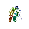
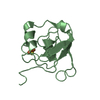
- PDB-6g61: Crystal structure of thioredoxin O1 from Arabidopsis thaliana in ... -
+ Open data

- Basic information
| Entry | Database: PDB / ID: 6g61 | ||||||
|---|---|---|---|---|---|---|---|
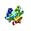
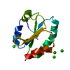
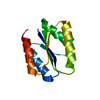
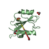
| Title | Crystal structure of thioredoxin O1 from Arabidopsis thaliana in oxidized state | ||||||
 Components Components | Thioredoxin O1, mitochondrial | ||||||
 Keywords Keywords |  OXIDOREDUCTASE / Thioredoxin OXIDOREDUCTASE / Thioredoxin | ||||||
| Function / homology |  Function and homology information Function and homology informationoxidoreductase activity, acting on a sulfur group of donors, disulfide as acceptor / mitochondrial matrix / endoplasmic reticulum / mitochondrion / cytosolSimilarity search - Function | ||||||
| Biological species |  Arabidopsis thaliana (thale cress) Arabidopsis thaliana (thale cress) | ||||||
| Method | X-RAY DIFFRACTION / SYNCHROTRON / Resolution: 1.8 Å | ||||||
 Authors Authors | Roret, T. / Didierjean, C. | ||||||
 Citation Citation | Journal: Antioxidants (Basel) / Year: 2018 Title: MitochondrialArabidopsis thalianaTRXo Isoforms Bind an Iron−Sulfur Cluster and Reduce NFU Proteins In Vitro. Authors: Zannini, F. / Roret, T. / Przybyla-Toscano, J. / Dhalleine, T. / Rouhier, N. / Couturier, J. | ||||||
| History |
|
- Structure visualization
| Structure viewer | Molecule: MolmilJmol/JSmol |
|---|
- Downloads & links
-Download
| PDBx/mmCIF format | 6g61.cif.gz | 75.5 KB | Display | PDBx/mmCIF format |
|---|---|---|---|---|
| PDB format | pdb6g61.ent.gz | 60.2 KB | Display | PDB format |
| PDBx/mmJSON format | 6g61.json.gz | Tree view | PDBx/mmJSON format | |
| Others |  Other downloads Other downloads |
-Validation report
| Arichive directory | https://data.pdbj.org/pub/pdb/validation_reports/g6/6g61ftp://data.pdbj.org/pub/pdb/validation_reports/g6/6g61 | HTTPS FTP |
|---|
-Related structure data


-Links
 PDBj
PDBj

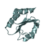
- Assembly
| Deposited unit | 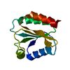
| ||||||||
|---|---|---|---|---|---|---|---|---|---|
| 1 |
| ||||||||
| Unit cell |
|
-Components
| #1: Protein | Mass: 14594.644 Da / Num. of mol.: 1 Source method: isolated from a genetically manipulated source Source: (gene. exp.) Arabidopsis thaliana (thale cress) / Gene: At2g35010, F19I3.24 / Production host:  Escherichia coli (E. coli) / References: UniProt: O64764 Escherichia coli (E. coli) / References: UniProt: O64764 |
|---|---|
| #2: Water | ChemComp-HOH / Water Mass: 18.015 Da / Num. of mol.: 130 / Source method: isolated from a natural source / Formula: H2O Mass: 18.015 Da / Num. of mol.: 130 / Source method: isolated from a natural source / Formula: H2O |
-Experimental details
-Experiment
| Experiment | Method: X-RAY DIFFRACTION / Number of used crystals: 1 |
|---|
- Sample preparation
| Crystal | Density Matthews: 1.98 Å3/Da / Density % sol: 37.92 % |
|---|---|
| Crystal grow | Temperature: 277 K / Method: microbatch / Details: 100 mM HEPES buffer (pH 7.5) 20% PEG 8000 |
-Data collection
| Diffraction | Mean temperature: 100 K |
|---|---|
| Diffraction source | Source: SYNCHROTRON / Site: ESRF  / Beamline: BM30A / Wavelength: 0.9799 Å / Beamline: BM30A / Wavelength: 0.9799 Å |
| Detector | Type: ADSC QUANTUM 315r / Detector: CCD / Date: Jun 23, 2017 |
| Radiation | Protocol: SINGLE WAVELENGTH / Monochromatic (M) / Laue (L): M / Scattering type: x-ray |
| Radiation wavelength | Wavelength: 0.9799 Å / Relative weight: 1 |
| Reflection | Resolution: 1.8→39.67 Å / Num. obs: 11316 / % possible obs: 100 % / Redundancy: 11.4 % / CC1/2: 0.996 / Rmerge(I) obs: 0.122 / Rpim(I) all: 0.049 / Rrim(I) all: 0.131 / Net I/σ(I): 13.1 |
| Reflection shell | Resolution: 1.8→1.84 Å / Redundancy: 7 % / Rmerge(I) obs: 0.242 / Mean I/σ(I) obs: 7 / Num. unique obs: 653 / CC1/2: 0.961 / Rpim(I) all: 0.145 / Rrim(I) all: 0.283 / % possible all: 100 |
- Processing
| Software |
| ||||||||||||||||||||||||||||||||||||||||||||||||||||||||||||||||||||||||||||||||||||||||||||||||||||||||||||||||||||||||||||||||||||||||||||||||||||||||||||||||||||||||||||||||||||||||||||||||||||||||
|---|---|---|---|---|---|---|---|---|---|---|---|---|---|---|---|---|---|---|---|---|---|---|---|---|---|---|---|---|---|---|---|---|---|---|---|---|---|---|---|---|---|---|---|---|---|---|---|---|---|---|---|---|---|---|---|---|---|---|---|---|---|---|---|---|---|---|---|---|---|---|---|---|---|---|---|---|---|---|---|---|---|---|---|---|---|---|---|---|---|---|---|---|---|---|---|---|---|---|---|---|---|---|---|---|---|---|---|---|---|---|---|---|---|---|---|---|---|---|---|---|---|---|---|---|---|---|---|---|---|---|---|---|---|---|---|---|---|---|---|---|---|---|---|---|---|---|---|---|---|---|---|---|---|---|---|---|---|---|---|---|---|---|---|---|---|---|---|---|---|---|---|---|---|---|---|---|---|---|---|---|---|---|---|---|---|---|---|---|---|---|---|---|---|---|---|---|---|---|---|---|---|
| Refinement | Resolution: 1.8→39.67 Å / SU ML: 0.22 / Cross valid method: FREE R-VALUE / σ(F): 1.34 / Phase error: 26.48
| ||||||||||||||||||||||||||||||||||||||||||||||||||||||||||||||||||||||||||||||||||||||||||||||||||||||||||||||||||||||||||||||||||||||||||||||||||||||||||||||||||||||||||||||||||||||||||||||||||||||||
| Solvent computation | Shrinkage radii: 0.9 Å / VDW probe radii: 1.11 Å | ||||||||||||||||||||||||||||||||||||||||||||||||||||||||||||||||||||||||||||||||||||||||||||||||||||||||||||||||||||||||||||||||||||||||||||||||||||||||||||||||||||||||||||||||||||||||||||||||||||||||
| Refinement step | Cycle: LAST / Resolution: 1.8→39.67 Å
| ||||||||||||||||||||||||||||||||||||||||||||||||||||||||||||||||||||||||||||||||||||||||||||||||||||||||||||||||||||||||||||||||||||||||||||||||||||||||||||||||||||||||||||||||||||||||||||||||||||||||
| Refine LS restraints |
| ||||||||||||||||||||||||||||||||||||||||||||||||||||||||||||||||||||||||||||||||||||||||||||||||||||||||||||||||||||||||||||||||||||||||||||||||||||||||||||||||||||||||||||||||||||||||||||||||||||||||
| LS refinement shell |
| ||||||||||||||||||||||||||||||||||||||||||||||||||||||||||||||||||||||||||||||||||||||||||||||||||||||||||||||||||||||||||||||||||||||||||||||||||||||||||||||||||||||||||||||||||||||||||||||||||||||||
| Refinement TLS params. | Method: refined / Refine-ID: X-RAY DIFFRACTION
| ||||||||||||||||||||||||||||||||||||||||||||||||||||||||||||||||||||||||||||||||||||||||||||||||||||||||||||||||||||||||||||||||||||||||||||||||||||||||||||||||||||||||||||||||||||||||||||||||||||||||
| Refinement TLS group |
|
