 Movie
Movie Controller

[English] 日本語
 Yorodumi
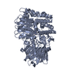
- PDB-6ftq: Crystal structure of human beta-ureidopropionase (beta-alanine sy... -
+ Open data

- Basic information
| Entry | Database: PDB / ID: 6ftq | ||||||
|---|---|---|---|---|---|---|---|
| Title | Crystal structure of human beta-ureidopropionase (beta-alanine synthase) - mutant T299C | ||||||
 Components Components | Beta-ureidopropionase | ||||||
 Keywords Keywords | HYDROLASE / pyrimidine degradation / N-carbamyl-beta-alanine amidohydrolase / 5-fluorouracil metabolizing enzyme | ||||||
| Function / homology |  Function and homology information Function and homology informationpyrimidine nucleoside catabolic process / beta-ureidopropionase / CMP catabolic process / UMP catabolic process / dCMP catabolic process / beta-alanine biosynthetic process via 3-ureidopropionate / beta-ureidopropionase activity / dUMP catabolic process / Pyrimidine catabolism / liver development ...pyrimidine nucleoside catabolic process / beta-ureidopropionase / CMP catabolic process / UMP catabolic process / dCMP catabolic process / beta-alanine biosynthetic process via 3-ureidopropionate / beta-ureidopropionase activity / dUMP catabolic process / Pyrimidine catabolism / liver development / protein homooligomerization / protein homotetramerization / in utero embryonic development / protein homodimerization activity / extracellular exosome / cytosolSimilarity search - Function | ||||||
| Biological species |  Homo sapiens (human) Homo sapiens (human) | ||||||
| Method | X-RAY DIFFRACTION / SYNCHROTRON / MOLECULAR REPLACEMENT / Resolution: 2.08 Å | ||||||
 Authors Authors | Dobritzsch, D. / Maurer, D. | ||||||
| Funding support |  Sweden, 1items Sweden, 1items
| ||||||
 Citation Citation | Journal: Biochem. J. / Year: 2018 Title: Crystal structure and pH-dependent allosteric regulation of human beta-ureidopropionase, an enzyme involved in anticancer drug metabolism. Authors: Maurer, D. / Lohkamp, B. / Krumpel, M. / Widersten, M. / Dobritzsch, D. | ||||||
| History |
|
- Structure visualization
| Structure viewer | Molecule: MolmilJmol/JSmol |
|---|
- Downloads & links
-Download
| PDBx/mmCIF format | 6ftq.cif.gz | 152 KB | Display | PDBx/mmCIF format |
|---|---|---|---|---|
| PDB format | pdb6ftq.ent.gz | 118.2 KB | Display | PDB format |
| PDBx/mmJSON format | 6ftq.json.gz | Tree view | PDBx/mmJSON format | |
| Others |  Other downloads Other downloads |
-Validation report
| Arichive directory | https://data.pdbj.org/pub/pdb/validation_reports/ft/6ftqftp://data.pdbj.org/pub/pdb/validation_reports/ft/6ftq | HTTPS FTP |
|---|
-Related structure data
| Related structure data | 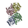 2vhhS S: Starting model for refinement |
|---|---|
| Similar structure data |


-Links
 PDBj
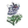
PDBj- Assembly
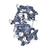
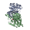
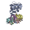
| Deposited unit | 
| ||||||||
|---|---|---|---|---|---|---|---|---|---|
| 1 | 
| ||||||||
| Unit cell |
| ||||||||
| Components on special symmetry positions |
|
-Components
| #1: Protein | / BUP-1 / Beta-alanine synthase / N-carbamoyl-beta-alanine amidohydrolase Mass: 43883.691 Da / Num. of mol.: 1 / Mutation: T299C Source method: isolated from a genetically manipulated source Details: the first 6 amino acids are a remainder of a cleaved His-tag C233 is oxidized to cysteine sulfinic acid T299 is mutated to cysteine Source: (gene. exp.) Homo sapiens (human) / Gene: UPB1, BUP1 / Plasmid: pET151/D-TOPO / Production host:  Escherichia coli BL21 (bacteria) / Variant (production host): AI / References: UniProt: Q9UBR1, beta-ureidopropionase Escherichia coli BL21 (bacteria) / Variant (production host): AI / References: UniProt: Q9UBR1, beta-ureidopropionase |
|---|---|
| #2: Water | ChemComp-HOH / Water Mass: 18.015 Da / Num. of mol.: 208 / Source method: isolated from a natural source / Formula: H2O Mass: 18.015 Da / Num. of mol.: 208 / Source method: isolated from a natural source / Formula: H2O |
-Experimental details
-Experiment
| Experiment | Method: X-RAY DIFFRACTION / Number of used crystals: 1 |
|---|
- Sample preparation
| Crystal | Density Matthews: 2.66 Å3/Da / Density % sol: 53.7 % |
|---|---|
| Crystal grow | Temperature: 293 K / Method: vapor diffusion, hanging drop / pH: 7.75 Details: 16% (w/v) PEG 3350 0.1 M HEPES pH 7.75 300 mM NaSCN 3 mg/ml enzyme variant |
-Data collection
| Diffraction | Mean temperature: 100 K |
|---|---|
| Diffraction source | Source: SYNCHROTRON / Site: Diamond  / Beamline: I03 / Wavelength: 0.9762 Å / Beamline: I03 / Wavelength: 0.9762 Å |
| Detector | Type: DECTRIS PILATUS3 6M / Detector: PIXEL / Date: Sep 25, 2017 |
| Radiation | Protocol: SINGLE WAVELENGTH / Monochromatic (M) / Laue (L): M / Scattering type: x-ray |
| Radiation wavelength | Wavelength: 0.9762 Å / Relative weight: 1 |
| Reflection | Resolution: 2.08→28.63 Å / Num. obs: 28448 / % possible obs: 99.6 % / Redundancy: 13.4 % / Biso Wilson estimate: 37.2 Å2 / CC1/2: 0.999 / Rmerge(I) obs: 0.126 / Rpim(I) all: 0.036 / Rrim(I) all: 0.131 / Net I/σ(I): 14.6 |
| Reflection shell | Resolution: 2.08→2.13 Å / Redundancy: 13 % / Rmerge(I) obs: 0.814 / Mean I/σ(I) obs: 3.3 / Num. unique obs: 2096 / CC1/2: 0.892 / Rpim(I) all: 0.232 / Rrim(I) all: 0.847 / % possible all: 95.1 |
- Processing
| Software |
| ||||||||||||||||||||||||||||||||||||||||||||||||||||||||||||||||||||||||||||||||||||||||||||||||||||||||||||||||||||||||||||||||||||||||||||||||||||||||||||||||||||||||||||||||||||||
|---|---|---|---|---|---|---|---|---|---|---|---|---|---|---|---|---|---|---|---|---|---|---|---|---|---|---|---|---|---|---|---|---|---|---|---|---|---|---|---|---|---|---|---|---|---|---|---|---|---|---|---|---|---|---|---|---|---|---|---|---|---|---|---|---|---|---|---|---|---|---|---|---|---|---|---|---|---|---|---|---|---|---|---|---|---|---|---|---|---|---|---|---|---|---|---|---|---|---|---|---|---|---|---|---|---|---|---|---|---|---|---|---|---|---|---|---|---|---|---|---|---|---|---|---|---|---|---|---|---|---|---|---|---|---|---|---|---|---|---|---|---|---|---|---|---|---|---|---|---|---|---|---|---|---|---|---|---|---|---|---|---|---|---|---|---|---|---|---|---|---|---|---|---|---|---|---|---|---|---|---|---|---|---|
| Refinement | Method to determine structure: MOLECULAR REPLACEMENT Starting model: 2VHH Resolution: 2.08→28.1 Å / Cor.coef. Fo:Fc: 0.956 / Cor.coef. Fo:Fc free: 0.948 / SU B: 6.925 / SU ML: 0.095 / Cross valid method: THROUGHOUT / ESU R: 0.159 / ESU R Free: 0.137 / Details: HYDROGENS HAVE BEEN ADDED IN THE RIDING POSITIONS
| ||||||||||||||||||||||||||||||||||||||||||||||||||||||||||||||||||||||||||||||||||||||||||||||||||||||||||||||||||||||||||||||||||||||||||||||||||||||||||||||||||||||||||||||||||||||
| Solvent computation | Ion probe radii: 0.8 Å / Shrinkage radii: 0.8 Å / VDW probe radii: 1.2 Å | ||||||||||||||||||||||||||||||||||||||||||||||||||||||||||||||||||||||||||||||||||||||||||||||||||||||||||||||||||||||||||||||||||||||||||||||||||||||||||||||||||||||||||||||||||||||
| Displacement parameters | Biso mean: 35.248 Å2
| ||||||||||||||||||||||||||||||||||||||||||||||||||||||||||||||||||||||||||||||||||||||||||||||||||||||||||||||||||||||||||||||||||||||||||||||||||||||||||||||||||||||||||||||||||||||
| Refinement step | Cycle: 1 / Resolution: 2.08→28.1 Å
| ||||||||||||||||||||||||||||||||||||||||||||||||||||||||||||||||||||||||||||||||||||||||||||||||||||||||||||||||||||||||||||||||||||||||||||||||||||||||||||||||||||||||||||||||||||||
| Refine LS restraints |
|