 Movie
Movie Controller

[English] 日本語
 Yorodumi
- PDB-2vhh: Crystal structure of a pyrimidine degrading enzyme from Drosophil... -
+ Open data

- Basic information
| Entry | Database: PDB / ID: 2vhh | ||||||
|---|---|---|---|---|---|---|---|
| Title | Crystal structure of a pyrimidine degrading enzyme from Drosophila melanogaster | ||||||
 Components Components | CG3027-PA | ||||||
 Keywords Keywords |  HYDROLASE HYDROLASE | ||||||
| Function / homology |  Function and homology informationPyrimidine catabolism / beta-ureidopropionase / pyrimidine nucleobase catabolic process / N-carbamoylputrescine amidase activity / beta-alanine biosynthetic process via 3-ureidopropionate / beta-ureidopropionase activity / putrescine biosynthetic process from arginine / Hydrolases; Acting on carbon-nitrogen bonds, other than peptide bonds; In linear amides / 'de novo' pyrimidine nucleobase biosynthetic process / cytoplasm Function and homology informationPyrimidine catabolism / beta-ureidopropionase / pyrimidine nucleobase catabolic process / N-carbamoylputrescine amidase activity / beta-alanine biosynthetic process via 3-ureidopropionate / beta-ureidopropionase activity / putrescine biosynthetic process from arginine / Hydrolases; Acting on carbon-nitrogen bonds, other than peptide bonds; In linear amides / 'de novo' pyrimidine nucleobase biosynthetic process / cytoplasmSimilarity search - Function | ||||||
| Biological species |  DROSOPHILA MELANOGASTER (fruit fly) DROSOPHILA MELANOGASTER (fruit fly) | ||||||
| Method | X-RAY DIFFRACTION / SYNCHROTRON / MOLECULAR REPLACEMENT / Resolution: 2.8 Å | ||||||
 Authors Authors | Lundgren, S. / Lohkamp, B. / Andersen, B. / Piskur, J. / Dobritzsch, D. | ||||||
 Citation Citation | Journal: J.Mol.Biol. / Year: 2008 Title: The Crystal Structure of Beta-Alanine Synthase from Drosophila Melanogaster Reveals a Homooctameric Helical Turn-Like Assembly. Authors: Lundgren, S. / Lohkamp, B. / Andersen, B. / Piskur, J. / Dobritzsch, D. | ||||||
| History |
|
- Structure visualization
| Structure viewer | Molecule: MolmilJmol/JSmol |
|---|
- Downloads & links
-Download
| PDBx/mmCIF format | 2vhh.cif.gz | 286.6 KB | Display | PDBx/mmCIF format |
|---|---|---|---|---|
| PDB format | pdb2vhh.ent.gz | 235.9 KB | Display | PDB format |
| PDBx/mmJSON format | 2vhh.json.gz | Tree view | PDBx/mmJSON format | |
| Others |  Other downloads Other downloads |
-Validation report
| Arichive directory | https://data.pdbj.org/pub/pdb/validation_reports/vh/2vhhftp://data.pdbj.org/pub/pdb/validation_reports/vh/2vhh | HTTPS FTP |
|---|
-Related structure data


-Links
 PDBj
PDBj- Assembly
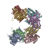
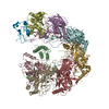
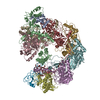
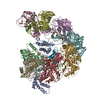
| Deposited unit | 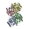
| |||||||||||||||||||||||||||||||||||||||||||||||||||||||||||||||||||||||||||||||||||||||||||||||||||||||||||||||||||||||||||||||||||||||||||||||||||||||||||||||||||||||||
|---|---|---|---|---|---|---|---|---|---|---|---|---|---|---|---|---|---|---|---|---|---|---|---|---|---|---|---|---|---|---|---|---|---|---|---|---|---|---|---|---|---|---|---|---|---|---|---|---|---|---|---|---|---|---|---|---|---|---|---|---|---|---|---|---|---|---|---|---|---|---|---|---|---|---|---|---|---|---|---|---|---|---|---|---|---|---|---|---|---|---|---|---|---|---|---|---|---|---|---|---|---|---|---|---|---|---|---|---|---|---|---|---|---|---|---|---|---|---|---|---|---|---|---|---|---|---|---|---|---|---|---|---|---|---|---|---|---|---|---|---|---|---|---|---|---|---|---|---|---|---|---|---|---|---|---|---|---|---|---|---|---|---|---|---|---|---|---|---|---|---|
| 1 | 
| |||||||||||||||||||||||||||||||||||||||||||||||||||||||||||||||||||||||||||||||||||||||||||||||||||||||||||||||||||||||||||||||||||||||||||||||||||||||||||||||||||||||||
| Unit cell |
| |||||||||||||||||||||||||||||||||||||||||||||||||||||||||||||||||||||||||||||||||||||||||||||||||||||||||||||||||||||||||||||||||||||||||||||||||||||||||||||||||||||||||
| Noncrystallographic symmetry (NCS) | NCS domain:
NCS domain segments: Refine code: 2
NCS ensembles :
NCS oper:
|
-Components
| #1: Protein | Mass: 46124.949 Da / Num. of mol.: 4 Source method: isolated from a genetically manipulated source Source: (gene. exp.) DROSOPHILA MELANOGASTER (fruit fly) / Production host:  ESCHERICHIA COLI (E. coli) / Strain (production host): BL21(DE3) / References: UniProt: Q9VI04, beta-ureidopropionase ESCHERICHIA COLI (E. coli) / Strain (production host): BL21(DE3) / References: UniProt: Q9VI04, beta-ureidopropionase |
|---|
-Experimental details
-Experiment
| Experiment | Method: X-RAY DIFFRACTION / Number of used crystals: 1 |
|---|
- Sample preparation
| Crystal | Density Matthews: 2.56 Å3/Da / Density % sol: 52 % / Description: NONE |
|---|---|
| Crystal grow | pH: 4.2 / Details: PEG 3350,PHOSPHATE/CITRATE PH 4.2, NACL |
-Data collection
| Diffraction | Mean temperature: 100 K |
|---|---|
| Diffraction source | Source: SYNCHROTRON / Site: ESRF  / Beamline: ID23-2 / Wavelength: 0.873 / Beamline: ID23-2 / Wavelength: 0.873 |
| Detector | Type: MARRESEARCH / Detector: CCD |
| Radiation | Protocol: SINGLE WAVELENGTH / Monochromatic (M) / Laue (L): M / Scattering type: x-ray |
| Radiation wavelength | Wavelength: 0.873 Å / Relative weight: 1 |
| Reflection | Resolution: 2.8→60 Å / Num. obs: 43922 / % possible obs: 95.6 % / Observed criterion σ(I): 2 / Redundancy: 2.7 % / Rmerge(I) obs: 0.12 / Net I/σ(I): 9.2 |
| Reflection shell | Resolution: 2.8→2.95 Å / Redundancy: 2.8 % / Rmerge(I) obs: 0.46 / Mean I/σ(I) obs: 2.3 / % possible all: 94.4 |
- Processing
| Software |
| ||||||||||||||||||||||||||||||||||||||||||||||||||||||||||||||||||||||||||||||||||||||||||||||||||||||||||||||||||||||||||||||||||||||||||||||||||||||||||||||||||||||||||||||||||||||
|---|---|---|---|---|---|---|---|---|---|---|---|---|---|---|---|---|---|---|---|---|---|---|---|---|---|---|---|---|---|---|---|---|---|---|---|---|---|---|---|---|---|---|---|---|---|---|---|---|---|---|---|---|---|---|---|---|---|---|---|---|---|---|---|---|---|---|---|---|---|---|---|---|---|---|---|---|---|---|---|---|---|---|---|---|---|---|---|---|---|---|---|---|---|---|---|---|---|---|---|---|---|---|---|---|---|---|---|---|---|---|---|---|---|---|---|---|---|---|---|---|---|---|---|---|---|---|---|---|---|---|---|---|---|---|---|---|---|---|---|---|---|---|---|---|---|---|---|---|---|---|---|---|---|---|---|---|---|---|---|---|---|---|---|---|---|---|---|---|---|---|---|---|---|---|---|---|---|---|---|---|---|---|---|
| Refinement | Method to determine structure: MOLECULAR REPLACEMENT / Resolution: 2.8→35 Å / Cor.coef. Fo:Fc: 0.908 / Cor.coef. Fo:Fc free: 0.877 / SU B: 14.178 / SU ML: 0.279 / Cross valid method: THROUGHOUT / ESU R Free: 0.395 / Stereochemistry target values: MAXIMUM LIKELIHOOD / Details: HYDROGENS HAVE BEEN ADDED IN THE RIDING POSITIONS
| ||||||||||||||||||||||||||||||||||||||||||||||||||||||||||||||||||||||||||||||||||||||||||||||||||||||||||||||||||||||||||||||||||||||||||||||||||||||||||||||||||||||||||||||||||||||
| Solvent computation | Ion probe radii: 0.8 Å / Shrinkage radii: 0.8 Å / VDW probe radii: 1.2 Å / Solvent model: MASK | ||||||||||||||||||||||||||||||||||||||||||||||||||||||||||||||||||||||||||||||||||||||||||||||||||||||||||||||||||||||||||||||||||||||||||||||||||||||||||||||||||||||||||||||||||||||
| Displacement parameters | Biso mean: 34.63 Å2
| ||||||||||||||||||||||||||||||||||||||||||||||||||||||||||||||||||||||||||||||||||||||||||||||||||||||||||||||||||||||||||||||||||||||||||||||||||||||||||||||||||||||||||||||||||||||
| Refinement step | Cycle: LAST / Resolution: 2.8→35 Å
| ||||||||||||||||||||||||||||||||||||||||||||||||||||||||||||||||||||||||||||||||||||||||||||||||||||||||||||||||||||||||||||||||||||||||||||||||||||||||||||||||||||||||||||||||||||||
| Refine LS restraints |
|