 Movie
Movie Controller

[English] 日本語
 Yorodumi
- PDB-6fp3: The crystal structure of EncM complexed with dioxygen under 5 bar... -
+ Open data

- Basic information
| Entry | Database: PDB / ID: 6fp3 | ||||||
|---|---|---|---|---|---|---|---|
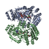
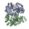
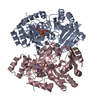
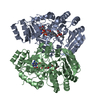
| Title | The crystal structure of EncM complexed with dioxygen under 5 bar of oxygen pressure. | ||||||
 Components Components | Putative FAD-dependent oxygenase EncM | ||||||
 Keywords Keywords |  FLAVOPROTEIN / nooxygenase / flavin-N5-oxide / FAD / EncM / oxygenating species / oxygen binding FLAVOPROTEIN / nooxygenase / flavin-N5-oxide / FAD / EncM / oxygenating species / oxygen binding | ||||||
| Function / homology |  Function and homology information Function and homology information | ||||||
| Biological species |  Streptomyces maritimus (bacteria) Streptomyces maritimus (bacteria) | ||||||
| Method | X-RAY DIFFRACTION / SYNCHROTRON / Resolution: 1.976 Å | ||||||
 Authors Authors | Saleem-Batcha, R. / Teufel, R. | ||||||
| Funding support |  Germany, 1items Germany, 1items
| ||||||
 Citation Citation | Journal: Proc. Natl. Acad. Sci. U.S.A. / Year: 2018 Title: Enzymatic control of dioxygen binding and functionalization of the flavin cofactor. Authors: Saleem-Batcha, R. / Stull, F. / Sanders, J.N. / Moore, B.S. / Palfey, B.A. / Houk, K.N. / Teufel, R. | ||||||
| History |
|
- Structure visualization
| Structure viewer | Molecule: MolmilJmol/JSmol |
|---|
- Downloads & links
-Download
| PDBx/mmCIF format | 6fp3.cif.gz | 379.7 KB | Display | PDBx/mmCIF format |
|---|---|---|---|---|
| PDB format | pdb6fp3.ent.gz | 308.4 KB | Display | PDB format |
| PDBx/mmJSON format | 6fp3.json.gz | Tree view | PDBx/mmJSON format | |
| Others |  Other downloads Other downloads |
-Validation report
| Arichive directory | https://data.pdbj.org/pub/pdb/validation_reports/fp/6fp3ftp://data.pdbj.org/pub/pdb/validation_reports/fp/6fp3 | HTTPS FTP |
|---|
-Related structure data
| Related structure data | 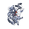 6foqC  6fowC 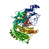 6fy8C 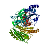 6fy9C  6fyaC 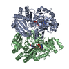 6fybC  6fycC  6fydC  6fyeC  6fyfC  6fygC C: citing same article ( |
|---|---|
| Similar structure data |

-Links
 PDBj
PDBj


- Assembly
| Deposited unit | 
| ||||||||||
|---|---|---|---|---|---|---|---|---|---|---|---|
| 1 | 
| ||||||||||
| 2 | 
| ||||||||||
| Unit cell |
|
-Components
| #1: Protein | Mass: 50074.371 Da / Num. of mol.: 4 Source method: isolated from a genetically manipulated source Source: (gene. exp.) Streptomyces maritimus (bacteria) / Gene: encM / Production host: Escherichia coli BL21(DE3) (bacteria) / References: UniProt: Q9KHK2#2: Chemical | ChemComp-FAD / Flavin adenine dinucleotide  Mass: 785.550 Da / Num. of mol.: 4 / Source method: obtained synthetically / Formula: C27H33N9O15P2 / Feature type: SUBJECT OF INVESTIGATION / Comment: FAD*YM Mass: 785.550 Da / Num. of mol.: 4 / Source method: obtained synthetically / Formula: C27H33N9O15P2 / Feature type: SUBJECT OF INVESTIGATION / Comment: FAD*YM#3: Chemical | ChemComp-OXY / Oxygen  Mass: 31.999 Da / Num. of mol.: 4 / Source method: obtained synthetically / Formula: O2 Mass: 31.999 Da / Num. of mol.: 4 / Source method: obtained synthetically / Formula: O2#4: Water | ChemComp-HOH / | Water Mass: 18.015 Da / Num. of mol.: 1018 / Source method: isolated from a natural source / Formula: H2O Mass: 18.015 Da / Num. of mol.: 1018 / Source method: isolated from a natural source / Formula: H2O |
|---|
-Experimental details
-Experiment
| Experiment | Method: X-RAY DIFFRACTION / Number of used crystals: 1 |
|---|
- Sample preparation
| Crystal | Density Matthews: 2.42 Å3/Da / Density % sol: 49.16 % |
|---|---|
| Crystal grow | Temperature: 295 K / Method: vapor diffusion, sitting drop / pH: 7.7 Details: 0.1M HEPES-Na, 0.2M calcium acetate, 20% PEG 3350, pH 7.5 |
-Data collection
| Diffraction | Mean temperature: 100 K |
|---|---|
| Diffraction source | Source: SYNCHROTRON / Site: SLS  / Beamline: X06SA / Wavelength: 1 Å / Beamline: X06SA / Wavelength: 1 Å |
| Detector | Type: DECTRIS EIGER X 16M / Detector: PIXEL / Date: Feb 17, 2017 |
| Radiation | Protocol: SINGLE WAVELENGTH / Monochromatic (M) / Laue (L): M / Scattering type: x-ray |
| Radiation wavelength | Wavelength: 1 Å / Relative weight: 1 |
| Reflection | Resolution: 1.975→48.91 Å / Num. obs: 131502 / % possible obs: 98.6 % / Redundancy: 6.4 % / Biso Wilson estimate: 30.1701402375 Å2 / Net I/σ(I): 7.3 |
| Reflection shell | Highest resolution: 1.975 Å |
- Processing
| Software |
| |||||||||||||||||||||||||||||||||||||||||||||||||||||||||||||||||||||||||||||||||||||||||||||||||||||||||||||||||||||||||||||||||||||||||||||||||||||||||||||||||||||||||||||||||||||||||||||||||||||||||||||||||||||||||
|---|---|---|---|---|---|---|---|---|---|---|---|---|---|---|---|---|---|---|---|---|---|---|---|---|---|---|---|---|---|---|---|---|---|---|---|---|---|---|---|---|---|---|---|---|---|---|---|---|---|---|---|---|---|---|---|---|---|---|---|---|---|---|---|---|---|---|---|---|---|---|---|---|---|---|---|---|---|---|---|---|---|---|---|---|---|---|---|---|---|---|---|---|---|---|---|---|---|---|---|---|---|---|---|---|---|---|---|---|---|---|---|---|---|---|---|---|---|---|---|---|---|---|---|---|---|---|---|---|---|---|---|---|---|---|---|---|---|---|---|---|---|---|---|---|---|---|---|---|---|---|---|---|---|---|---|---|---|---|---|---|---|---|---|---|---|---|---|---|---|---|---|---|---|---|---|---|---|---|---|---|---|---|---|---|---|---|---|---|---|---|---|---|---|---|---|---|---|---|---|---|---|---|---|---|---|---|---|---|---|---|---|---|---|---|---|---|---|---|
| Refinement | Resolution: 1.976→48.91 Å / SU ML: 0.22 / Cross valid method: NONE / σ(F): 1.33 / Phase error: 21.76 / Stereochemistry target values: ML
| |||||||||||||||||||||||||||||||||||||||||||||||||||||||||||||||||||||||||||||||||||||||||||||||||||||||||||||||||||||||||||||||||||||||||||||||||||||||||||||||||||||||||||||||||||||||||||||||||||||||||||||||||||||||||
| Solvent computation | Shrinkage radii: 0.9 Å / VDW probe radii: 1.11 Å / Solvent model: FLAT BULK SOLVENT MODEL | |||||||||||||||||||||||||||||||||||||||||||||||||||||||||||||||||||||||||||||||||||||||||||||||||||||||||||||||||||||||||||||||||||||||||||||||||||||||||||||||||||||||||||||||||||||||||||||||||||||||||||||||||||||||||
| Refinement step | Cycle: LAST / Resolution: 1.976→48.91 Å
| |||||||||||||||||||||||||||||||||||||||||||||||||||||||||||||||||||||||||||||||||||||||||||||||||||||||||||||||||||||||||||||||||||||||||||||||||||||||||||||||||||||||||||||||||||||||||||||||||||||||||||||||||||||||||
| Refine LS restraints |
| |||||||||||||||||||||||||||||||||||||||||||||||||||||||||||||||||||||||||||||||||||||||||||||||||||||||||||||||||||||||||||||||||||||||||||||||||||||||||||||||||||||||||||||||||||||||||||||||||||||||||||||||||||||||||
| LS refinement shell |
|
