 Movie
Movie Controller


+ Open data

- Basic information
| Entry | Database: PDB / ID: 4xlo | ||||||
|---|---|---|---|---|---|---|---|
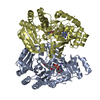
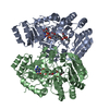
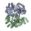
| Title | Crystal Structure of EncM (crystallized with 4 mM NADPH) | ||||||
 Components Components | FAD-dependent oxygenase EncM | ||||||
 Keywords Keywords |  OXIDOREDUCTASE / Flavoenzyme / NADPH / vanillyl-alcohol oxidase/p-cresol methylhydroxylase fold / oxygenase OXIDOREDUCTASE / Flavoenzyme / NADPH / vanillyl-alcohol oxidase/p-cresol methylhydroxylase fold / oxygenase | ||||||
| Function / homology |  Function and homology information Function and homology information | ||||||
| Biological species |  Streptomyces maritimus (bacteria) Streptomyces maritimus (bacteria) | ||||||
| Method | X-RAY DIFFRACTION / SYNCHROTRON / MOLECULAR REPLACEMENT / Resolution: 1.67 Å | ||||||
 Authors Authors | Teufel, R. | ||||||
| Funding support |  United States, 1items United States, 1items
| ||||||
 Citation Citation | Journal: Nature / Year: 2013 Title: Flavin-mediated dual oxidation controls an enzymatic Favorskii-type rearrangement. Authors: Teufel, R. / Miyanaga, A. / Michaudel, Q. / Stull, F. / Louie, G. / Noel, J.P. / Baran, P.S. / Palfey, B. / Moore, B.S. #1: Journal: Nature / Year: 2013Title: Flavin-mediated dual oxidation controls an enzymatic Favorskii-type rearrangement Authors: Teufel, R. / Miyanaga, A. / Michaudel, Q. / Stull, F. / Louie, G. / Noel, J.P. / Baran, P.S. / Palfey, B. / Moore, B.S. | ||||||
| History |
|
- Structure visualization
| Structure viewer | Molecule: MolmilJmol/JSmol |
|---|
- Downloads & links
-Download
| PDBx/mmCIF format | 4xlo.cif.gz | 413.7 KB | Display | PDBx/mmCIF format |
|---|---|---|---|---|
| PDB format | pdb4xlo.ent.gz | 333.5 KB | Display | PDB format |
| PDBx/mmJSON format | 4xlo.json.gz | Tree view | PDBx/mmJSON format | |
| Others |  Other downloads Other downloads |
-Validation report
| Arichive directory | https://data.pdbj.org/pub/pdb/validation_reports/xl/4xloftp://data.pdbj.org/pub/pdb/validation_reports/xl/4xlo | HTTPS FTP |
|---|
-Related structure data
| Related structure data | 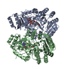 3w8wC 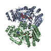 3w8xC  3w8zC 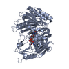 2bvgS C: citing same article ( S: Starting model for refinement |
|---|---|
| Similar structure data |


-Links
 PDBj
PDBj
- Assembly
| Deposited unit | 
| ||||||||
|---|---|---|---|---|---|---|---|---|---|
| 1 | 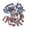
| ||||||||
| 2 | 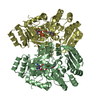
| ||||||||
| Unit cell |
| ||||||||
| Components on special symmetry positions |
|
-Components
| #1: Protein | Mass: 50074.371 Da / Num. of mol.: 4 Source method: isolated from a genetically manipulated source Source: (gene. exp.) Streptomyces maritimus (bacteria) / Gene: encM / Production host: Escherichia coli BL21(DE3) (bacteria) / References: UniProt: Q9KHK2#2: Chemical | ChemComp-FAD / Flavin adenine dinucleotide  Mass: 785.550 Da / Num. of mol.: 4 Mass: 785.550 Da / Num. of mol.: 4Source method: isolated from a genetically manipulated source Formula: C27H33N9O15P2 / Comment: FAD*YM #3: Water | ChemComp-HOH / | Water Mass: 18.015 Da / Num. of mol.: 2120 / Source method: isolated from a natural source / Formula: H2O Mass: 18.015 Da / Num. of mol.: 2120 / Source method: isolated from a natural source / Formula: H2O |
|---|
-Experimental details
-Experiment
| Experiment | Method: X-RAY DIFFRACTION |
|---|
- Sample preparation
| Crystal | Density Matthews: 2.39 Å3/Da / Density % sol: 48.44 % |
|---|---|
| Crystal grow | Temperature: 277.15 K / Method: vapor diffusion, hanging drop Details: 5 mg/mL EncM in 4 mM NADPH, 5 mM TES sodium, pH 7.7, 10% v/v glycerol, reservoir: 0.1 M HEPES sodium, pH 7.5, 0.2 M calcium acetate, 20% w/v PEG3350 PH range: 7.5-7.7 |
-Data collection
| Diffraction | Mean temperature: 100 K |
|---|---|
| Diffraction source | Source: SYNCHROTRON / Site: ALS / Beamline: 8.2.1 / Wavelength: 1 Å |
| Detector | Type: ADSC QUANTUM 315r / Detector: CCD / Date: Jul 10, 2012 |
| Radiation | Monochromator: double crystal Si(111) / Protocol: SINGLE WAVELENGTH / Monochromatic (M) / Laue (L): M / Scattering type: x-ray |
| Radiation wavelength | Wavelength: 1 Å / Relative weight: 1 |
| Reflection | Resolution: 1.67→29.69 Å / Num. obs: 219275 / % possible obs: 98.35 % / Redundancy: 6.6 % / Rmerge(I) obs: 0.155 / Net I/σ(I): 28.8 |
- Processing
| Software |
| |||||||||||||||||||||||||||||||||||||||||||||
|---|---|---|---|---|---|---|---|---|---|---|---|---|---|---|---|---|---|---|---|---|---|---|---|---|---|---|---|---|---|---|---|---|---|---|---|---|---|---|---|---|---|---|---|---|---|---|
| Refinement | Method to determine structure: MOLECULAR REPLACEMENT Starting model: PDB entry 2BVG Resolution: 1.67→29.69 Å / Cor.coef. Fo:Fc: 0.964 / Cor.coef. Fo:Fc free: 0.947 / SU B: 2.669 / SU ML: 0.085 / Cross valid method: THROUGHOUT / σ(F): 0 / ESU R: 0.109 / ESU R Free: 0.112 / Stereochemistry target values: MAXIMUM LIKELIHOOD / Details: HYDROGENS HAVE BEEN USED IF PRESENT IN THE INPUT
| |||||||||||||||||||||||||||||||||||||||||||||
| Solvent computation | Ion probe radii: 0.8 Å / Shrinkage radii: 0.8 Å / VDW probe radii: 1.2 Å / Solvent model: MASK | |||||||||||||||||||||||||||||||||||||||||||||
| Displacement parameters | Biso max: 104.84 Å2 / Biso mean: 23.32 Å2 / Biso min: 8.45 Å2
| |||||||||||||||||||||||||||||||||||||||||||||
| Refinement step | Cycle: final / Resolution: 1.67→29.69 Å
| |||||||||||||||||||||||||||||||||||||||||||||
| Refine LS restraints |
| |||||||||||||||||||||||||||||||||||||||||||||
| LS refinement shell | Resolution: 1.671→1.714 Å / Total num. of bins used: 20
|
