 Movie
Movie Controller

[English] 日本語
 Yorodumi
- PDB-6fl5: Structure of human SHMT1-H135N-R137A-E168N mutant at 3.6 Ang. res... -
+ Open data

- Basic information
| Entry | Database: PDB / ID: 6fl5 | |||||||||||||||
|---|---|---|---|---|---|---|---|---|---|---|---|---|---|---|---|---|
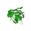
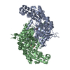
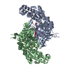
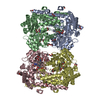
| Title | Structure of human SHMT1-H135N-R137A-E168N mutant at 3.6 Ang. resolution | |||||||||||||||
 Components Components | Serine hydroxymethyltransferase, cytosolic | |||||||||||||||
 Keywords Keywords | TRANSFERASE / serine hydroxymethyltransferase / Interface / Tetramer / OCM / serine / THF / glicine / TCA | |||||||||||||||
| Function / homology |  Function and homology information Function and homology informationcellular response to tetrahydrofolate / Carnitine synthesis / purine nucleobase biosynthetic process / L-serine metabolic process / glycine metabolic process / serine binding / glycine hydroxymethyltransferase / glycine hydroxymethyltransferase activity / glycine biosynthetic process from serine / L-serine catabolic process ...cellular response to tetrahydrofolate / Carnitine synthesis / purine nucleobase biosynthetic process / L-serine metabolic process / glycine metabolic process / serine binding / glycine hydroxymethyltransferase / glycine hydroxymethyltransferase activity / glycine biosynthetic process from serine / L-serine catabolic process / Metabolism of folate and pterines / tetrahydrofolate metabolic process / tetrahydrofolate interconversion / dTMP biosynthetic process / cobalt ion binding / folic acid metabolic process / small molecule binding / mRNA regulatory element binding translation repressor activity / cellular response to leukemia inhibitory factor / mRNA 5'-UTR binding / pyridoxal phosphate binding / protein homotetramerization / negative regulation of translation / protein homodimerization activity / mitochondrion / extracellular exosome / zinc ion binding / nucleoplasm / identical protein binding / cytosol / cytoplasmSimilarity search - Function | |||||||||||||||
| Biological species |  Homo sapiens (human) Homo sapiens (human) | |||||||||||||||
| Method | X-RAY DIFFRACTION / SYNCHROTRON / MOLECULAR REPLACEMENT / molecular replacement / Resolution: 3.6 Å | |||||||||||||||
 Authors Authors | Giardina, G. / Cutruzzola, F. / Lucchi, R. | |||||||||||||||
| Funding support |  Italy, 4items Italy, 4items
| |||||||||||||||
 Citation Citation | Journal: FEBS J. / Year: 2018 Title: The catalytic activity of serine hydroxymethyltransferase is essential for de novo nuclear dTMP synthesis in lung cancer cells. Authors: Giardina, G. / Paone, A. / Tramonti, A. / Lucchi, R. / Marani, M. / Magnifico, M.C. / Bouzidi, A. / Pontecorvi, V. / Guiducci, G. / Zamparelli, C. / Rinaldo, S. / Paiardini, A. / ...Authors: Giardina, G. / Paone, A. / Tramonti, A. / Lucchi, R. / Marani, M. / Magnifico, M.C. / Bouzidi, A. / Pontecorvi, V. / Guiducci, G. / Zamparelli, C. / Rinaldo, S. / Paiardini, A. / Contestabile, R. / Cutruzzola, F. | |||||||||||||||
| History |
|
- Structure visualization
| Structure viewer | Molecule: MolmilJmol/JSmol |
|---|
- Downloads & links
-Download
| PDBx/mmCIF format | 6fl5.cif.gz | 358.7 KB | Display | PDBx/mmCIF format |
|---|---|---|---|---|
| PDB format | pdb6fl5.ent.gz | 292.1 KB | Display | PDB format |
| PDBx/mmJSON format | 6fl5.json.gz | Tree view | PDBx/mmJSON format | |
| Others |  Other downloads Other downloads |
-Validation report
| Arichive directory | https://data.pdbj.org/pub/pdb/validation_reports/fl/6fl5ftp://data.pdbj.org/pub/pdb/validation_reports/fl/6fl5 | HTTPS FTP |
|---|
-Related structure data
| Related structure data | 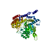 1bj4S S: Starting model for refinement |
|---|---|
| Similar structure data |
-Links
 PDBj
PDBj
- Assembly
| Deposited unit | 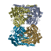
| ||||||||||||||||||||||||||||||||||||||||||||||||||||||||||||||||||||||||||||||||||||||||||||||||||||||||||||||||||||||||||||||||||||||||||||||||||||||||||||||||||||||||||||||||||||||||||||||||||||||||||||||||||||||||||||||||||||
|---|---|---|---|---|---|---|---|---|---|---|---|---|---|---|---|---|---|---|---|---|---|---|---|---|---|---|---|---|---|---|---|---|---|---|---|---|---|---|---|---|---|---|---|---|---|---|---|---|---|---|---|---|---|---|---|---|---|---|---|---|---|---|---|---|---|---|---|---|---|---|---|---|---|---|---|---|---|---|---|---|---|---|---|---|---|---|---|---|---|---|---|---|---|---|---|---|---|---|---|---|---|---|---|---|---|---|---|---|---|---|---|---|---|---|---|---|---|---|---|---|---|---|---|---|---|---|---|---|---|---|---|---|---|---|---|---|---|---|---|---|---|---|---|---|---|---|---|---|---|---|---|---|---|---|---|---|---|---|---|---|---|---|---|---|---|---|---|---|---|---|---|---|---|---|---|---|---|---|---|---|---|---|---|---|---|---|---|---|---|---|---|---|---|---|---|---|---|---|---|---|---|---|---|---|---|---|---|---|---|---|---|---|---|---|---|---|---|---|---|---|---|---|---|---|---|---|---|---|---|
| 1 |
| ||||||||||||||||||||||||||||||||||||||||||||||||||||||||||||||||||||||||||||||||||||||||||||||||||||||||||||||||||||||||||||||||||||||||||||||||||||||||||||||||||||||||||||||||||||||||||||||||||||||||||||||||||||||||||||||||||||
| Unit cell |
| ||||||||||||||||||||||||||||||||||||||||||||||||||||||||||||||||||||||||||||||||||||||||||||||||||||||||||||||||||||||||||||||||||||||||||||||||||||||||||||||||||||||||||||||||||||||||||||||||||||||||||||||||||||||||||||||||||||
| Noncrystallographic symmetry (NCS) | NCS domain:
NCS domain segments: Component-ID: 1 / Refine code: 1
NCS ensembles :
NCS oper:
|
-Components
| #1: Protein | / SHMT / Glycine hydroxymethyltransferase / Serine methylase Mass: 51695.797 Da / Num. of mol.: 4 / Mutation: H135N, R137A, E168N Source method: isolated from a genetically manipulated source Source: (gene. exp.) Homo sapiens (human) / Gene: SHMT1 / Plasmid: pET28b / Production host:  Escherichia coli BL21(DE3) (bacteria) Escherichia coli BL21(DE3) (bacteria)References: UniProt: P34896, glycine hydroxymethyltransferase#2: Chemical | ChemComp-PLP / Pyridoxal phosphate  Mass: 247.142 Da / Num. of mol.: 4 / Source method: obtained synthetically / Formula: C8H10NO6P Mass: 247.142 Da / Num. of mol.: 4 / Source method: obtained synthetically / Formula: C8H10NO6P#3: Chemical | ChemComp-CL / Chloride  Mass: 35.453 Da / Num. of mol.: 4 / Source method: obtained synthetically / Formula: Cl Mass: 35.453 Da / Num. of mol.: 4 / Source method: obtained synthetically / Formula: Cl |
|---|
-Experimental details
-Experiment
| Experiment | Method: X-RAY DIFFRACTION / Number of used crystals: 1 |
|---|
- Sample preparation
| Crystal | Density Matthews: 3.17 Å3/Da / Density % sol: 61.24 % / Description: rod like crystals |
|---|---|
| Crystal grow | Temperature: 294 K / Method: vapor diffusion, hanging drop / pH: 6.5 Details: 2 microL of 80microM protein solution in: 20 mM Hepes pH7.2, 250 mM NaCl 5% glycerol + 2 microL of reservoir:0.1 M Na Cacodilate pH6.5 - 1M Na citrate |
-Data collection
| Diffraction | Mean temperature: 100 K | ||||||||||||||||||||||||
|---|---|---|---|---|---|---|---|---|---|---|---|---|---|---|---|---|---|---|---|---|---|---|---|---|---|
| Diffraction source | Source: SYNCHROTRON / Site: ESRF  / Beamline: BM30A / Wavelength: 0.9677 Å / Beamline: BM30A / Wavelength: 0.9677 Å | ||||||||||||||||||||||||
| Detector | Type: DECTRIS EIGER R 4M / Detector: PIXEL / Date: Mar 10, 2017 / Details: CRL | ||||||||||||||||||||||||
| Radiation | Monochromator: C(110) / Protocol: SINGLE WAVELENGTH / Monochromatic (M) / Laue (L): M / Scattering type: x-ray | ||||||||||||||||||||||||
| Radiation wavelength | Wavelength: 0.9677 Å / Relative weight: 1 | ||||||||||||||||||||||||
| Reflection | Resolution: 3.6→56.61 Å / Num. obs: 30938 / % possible obs: 98.2 % / Redundancy: 6.3 % / CC1/2: 0.973 / Rmerge(I) obs: 0.312 / Rpim(I) all: 0.131 / Rrim(I) all: 0.34 / Net I/σ(I): 5.6 | ||||||||||||||||||||||||
| Reflection shell | Diffraction-ID: 1
|
-Phasing
| Phasing | Method: molecular replacement | ||||||
|---|---|---|---|---|---|---|---|
| Phasing MR | R rigid body: 0.57
|
- Processing
| Software |
| ||||||||||||||||||||||||||||||||||||||||||||||||||||||||||||
|---|---|---|---|---|---|---|---|---|---|---|---|---|---|---|---|---|---|---|---|---|---|---|---|---|---|---|---|---|---|---|---|---|---|---|---|---|---|---|---|---|---|---|---|---|---|---|---|---|---|---|---|---|---|---|---|---|---|---|---|---|---|
| Refinement | Method to determine structure: MOLECULAR REPLACEMENT Starting model: 1BJ4 Resolution: 3.6→50.01 Å / Cor.coef. Fo:Fc: 0.865 / Cor.coef. Fo:Fc free: 0.83 / SU B: 44.648 / SU ML: 0.62 / Cross valid method: THROUGHOUT / σ(F): 0 / ESU R Free: 0.716 Details: HYDROGENS HAVE BEEN ADDED IN THE RIDING POSITIONS U VALUES : REFINED INDIVIDUALLY
| ||||||||||||||||||||||||||||||||||||||||||||||||||||||||||||
| Solvent computation | Ion probe radii: 0.8 Å / Shrinkage radii: 0.8 Å / VDW probe radii: 1.2 Å | ||||||||||||||||||||||||||||||||||||||||||||||||||||||||||||
| Displacement parameters | Biso max: 160.73 Å2 / Biso mean: 74.364 Å2 / Biso min: 19.04 Å2
| ||||||||||||||||||||||||||||||||||||||||||||||||||||||||||||
| Refinement step | Cycle: final / Resolution: 3.6→50.01 Å
| ||||||||||||||||||||||||||||||||||||||||||||||||||||||||||||
| Refine LS restraints |
| ||||||||||||||||||||||||||||||||||||||||||||||||||||||||||||
| Refine LS restraints NCS | Dom-ID: 1 / Refine-ID: X-RAY DIFFRACTION / Type: TIGHT THERMAL / Weight position: 0.87
| ||||||||||||||||||||||||||||||||||||||||||||||||||||||||||||
| LS refinement shell | Resolution: 3.6→3.693 Å / Rfactor Rfree error: 0 / Total num. of bins used: 20
|
