 Movie
Movie Controller

[English] 日本語
 Yorodumi
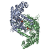
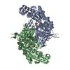
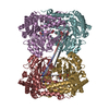
- PDB-1rvy: E75Q MUTANT OF RABBIT CYTOSOLIC SERINE HYDROXYMETHYLTRANSFERASE, ... -
+ Open data

- Basic information
| Entry | Database: PDB / ID: 1rvy | ||||||
|---|---|---|---|---|---|---|---|
| Title | E75Q MUTANT OF RABBIT CYTOSOLIC SERINE HYDROXYMETHYLTRANSFERASE, COMPLEX WITH GLYCINE | ||||||
 Components Components | Serine hydroxymethyltransferase, cytosolic | ||||||
 Keywords Keywords | HYDROLASE / ONE CARBON METABOLISM | ||||||
| Function / homology |  Function and homology information Function and homology informationcellular response to tetrahydrofolate / purine nucleobase biosynthetic process / L-serine metabolic process / glycine metabolic process / serine binding / glycine hydroxymethyltransferase / glycine hydroxymethyltransferase activity / glycine biosynthetic process from serine / L-serine catabolic process / tetrahydrofolate metabolic process ...cellular response to tetrahydrofolate / purine nucleobase biosynthetic process / L-serine metabolic process / glycine metabolic process / serine binding / glycine hydroxymethyltransferase / glycine hydroxymethyltransferase activity / glycine biosynthetic process from serine / L-serine catabolic process / tetrahydrofolate metabolic process / tetrahydrofolate interconversion / folic acid metabolic process / mRNA regulatory element binding translation repressor activity / mRNA 5'-UTR binding / pyridoxal phosphate binding / protein homotetramerization / protein homodimerization activity / nucleoplasm / cytosolSimilarity search - Function | ||||||
| Biological species |  Oryctolagus cuniculus (rabbit) Oryctolagus cuniculus (rabbit) | ||||||
| Method | X-RAY DIFFRACTION / Rigid body refinement of unliganded E75Q rcSHMT structure / Resolution: 2.9 Å | ||||||
 Authors Authors | Szebenyi, D.M. / Musayev, F.N. / Di Salvo, M.L. / Safo, M.K. / Schirch, V. | ||||||
 Citation Citation | Journal: Biochemistry / Year: 2004 Title: Serine Hydroxymethyltransferase: Role of Glu75 and Evidence that Serine Is Cleaved by a Retroaldol Mechanism. Authors: Szebenyi, D.M. / Musayev, F.N. / Di Salvo, M.L. / Safo, M.K. / Schirch, V. | ||||||
| History |
|
- Structure visualization
| Structure viewer | Molecule: MolmilJmol/JSmol |
|---|
- Downloads & links
-Download
| PDBx/mmCIF format | 1rvy.cif.gz | 191.6 KB | Display | PDBx/mmCIF format |
|---|---|---|---|---|
| PDB format | pdb1rvy.ent.gz | 152.3 KB | Display | PDB format |
| PDBx/mmJSON format | 1rvy.json.gz | Tree view | PDBx/mmJSON format | |
| Others |  Other downloads Other downloads |
-Validation report
| Arichive directory | https://data.pdbj.org/pub/pdb/validation_reports/rv/1rvyftp://data.pdbj.org/pub/pdb/validation_reports/rv/1rvy | HTTPS FTP |
|---|
-Related structure data
| Related structure data |  1rv3C 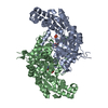 1rv4C  1rvuSC C: citing same article ( S: Starting model for refinement |
|---|---|
| Similar structure data |


-Links
 PDBj
PDBj- Assembly
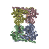
| Deposited unit | 
| ||||||||
|---|---|---|---|---|---|---|---|---|---|
| 1 | 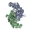
| ||||||||
| Unit cell |
| ||||||||
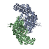
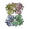
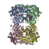
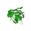
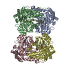
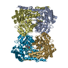
| Details | The asymmetric unit contains a homodimer. The biological assembly is a tetramer consisting of a pair of dimers. The second dimer in the tetramer is generated by the operation: y,x,1-z. |
-Components
| #1: Protein | / Serine methylase / Glycine hydroxymethyltransferase / SHMT Mass: 52915.129 Da / Num. of mol.: 2 / Mutation: E75Q Source method: isolated from a genetically manipulated source Source: (gene. exp.) Oryctolagus cuniculus (rabbit) / Gene: SHMT1 / Plasmid: pET22b / Production host:  Escherichia coli (E. coli) Escherichia coli (E. coli)References: UniProt: P07511, glycine hydroxymethyltransferase#2: Chemical | ChemComp-PLG / |   Mass: 306.209 Da / Num. of mol.: 1 / Source method: obtained synthetically / Formula: C10H15N2O7P Mass: 306.209 Da / Num. of mol.: 1 / Source method: obtained synthetically / Formula: C10H15N2O7P#3: Chemical | ChemComp-PO4 / | Phosphate  Mass: 94.971 Da / Num. of mol.: 1 / Source method: obtained synthetically / Formula: PO4 Mass: 94.971 Da / Num. of mol.: 1 / Source method: obtained synthetically / Formula: PO4#4: Chemical | ChemComp-PLP / | Pyridoxal phosphate  Mass: 247.142 Da / Num. of mol.: 1 / Source method: obtained synthetically / Formula: C8H10NO6P Mass: 247.142 Da / Num. of mol.: 1 / Source method: obtained synthetically / Formula: C8H10NO6P#5: Water | ChemComp-HOH / | Water Mass: 18.015 Da / Num. of mol.: 102 / Source method: isolated from a natural source / Formula: H2O Mass: 18.015 Da / Num. of mol.: 102 / Source method: isolated from a natural source / Formula: H2O |
|---|
-Experimental details
-Experiment
| Experiment | Method: X-RAY DIFFRACTION / Number of used crystals: 1 |
|---|
- Sample preparation
| Crystal | Density Matthews: 2.43 Å3/Da / Density % sol: 49.38 % |
|---|---|
| Crystal grow | Temperature: 298 K / Method: vapor diffusion, hanging drop / pH: 6.4 Details: PEG 4000, KCl, KMES, pH 6.4, VAPOR DIFFUSION, HANGING DROP, temperature 298.K |
-Data collection
| Diffraction | Mean temperature: 100 K |
|---|---|
| Diffraction source | Source: ROTATING ANODE / Type: RIGAKU / Wavelength: 1.5418 Å |
| Detector | Type: RIGAKU RAXIS II / Detector: IMAGE PLATE / Date: Mar 3, 2003 / Details: Mirrors |
| Radiation | Monochromator: GRAPHITE / Protocol: SINGLE WAVELENGTH / Monochromatic (M) / Laue (L): M / Scattering type: x-ray |
| Radiation wavelength | Wavelength: 1.5418 Å / Relative weight: 1 |
| Reflection | Resolution: 2.9→27.2 Å / Num. all: 22342 / Num. obs: 22342 / % possible obs: 93.8 % / Observed criterion σ(F): 0 / Observed criterion σ(I): 0 / Redundancy: 5.8 % / Biso Wilson estimate: 28.3 Å2 / Rsym value: 0.188 / Net I/σ(I): 3.8 |
| Reflection shell | Resolution: 2.9→3.06 Å / Redundancy: 5.7 % / Mean I/σ(I) obs: 1.1 / Num. unique all: 3388 / Rsym value: 0.626 / % possible all: 99.9 |
- Processing
| Software |
| |||||||||||||||||||||||||
|---|---|---|---|---|---|---|---|---|---|---|---|---|---|---|---|---|---|---|---|---|---|---|---|---|---|---|
| Refinement | Method to determine structure: Rigid body refinement of unliganded E75Q rcSHMT structure Starting model: PDB entry 1RVU Resolution: 2.9→27.16 Å / Rfactor Rfree error: 0.008 / Data cutoff high absF: 4024355.57 / Data cutoff low absF: 0 / Isotropic thermal model: GROUP / Cross valid method: THROUGHOUT / σ(F): 0 / Stereochemistry target values: Engh & Huber Details: Solvent molecules carried over from (higher resolution) unliganded structure
| |||||||||||||||||||||||||
| Solvent computation | Solvent model: FLAT MODEL / Bsol: 10.6844 Å2 / ksol: 0.26216 e/Å3 | |||||||||||||||||||||||||
| Displacement parameters | Biso mean: 33.4 Å2
| |||||||||||||||||||||||||
| Refine analyze |
| |||||||||||||||||||||||||
| Refinement step | Cycle: LAST / Resolution: 2.9→27.16 Å
| |||||||||||||||||||||||||
| Refine LS restraints |
| |||||||||||||||||||||||||
| LS refinement shell | Resolution: 2.9→3.08 Å / Rfactor Rfree error: 0.026 / Total num. of bins used: 6
| |||||||||||||||||||||||||
| Xplor file |
|
