 Movie
Movie Controller


+ Open data

- Basic information
| Entry | Database: PDB / ID: 6fdy | ||||||
|---|---|---|---|---|---|---|---|
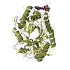
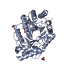
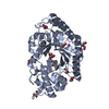
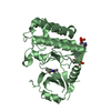
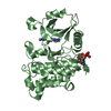
| Title | Unc-51-Like Kinase 3 (ULK3) In Complex With Bosutinib | ||||||
 Components Components | Serine/threonine-protein kinase ULK3 | ||||||
 Keywords Keywords |  TRANSFERASE / Kinase Inhibitor DFG-in TRANSFERASE / Kinase Inhibitor DFG-in | ||||||
| Function / homology |  Function and homology information Function and homology informationciliary tip / phagophore assembly site membrane / piecemeal microautophagy of the nucleus / fibroblast activation / positive regulation of smoothened signaling pathway / phagophore assembly site / reticulophagy / smoothened signaling pathway / response to starvation / autophagosome assembly ...ciliary tip / phagophore assembly site membrane / piecemeal microautophagy of the nucleus / fibroblast activation / positive regulation of smoothened signaling pathway / phagophore assembly site / reticulophagy / smoothened signaling pathway / response to starvation / autophagosome assembly / autophagosome / negative regulation of smoothened signaling pathway / regulation of autophagy / Hedgehog 'on' state / autophagy / cellular senescence / protein autophosphorylation / non-specific serine/threonine protein kinase / protein serine kinase activity / protein serine/threonine kinase activity / ATP binding / cytosol / cytoplasmSimilarity search - Function | ||||||
| Biological species |  Homo sapiens (human) Homo sapiens (human) | ||||||
| Method | X-RAY DIFFRACTION / SYNCHROTRON / MOLECULAR REPLACEMENT / Resolution: 1.7 Å | ||||||
 Authors Authors | Mathea, S. / Salah, E. / Moroglu, M. / Scorah, A. / Krojer, T. / von Delft, F. / Arrowsmith, C.H. / Edwards, A.M. / Bountra, C. / Huber, K. / Knapp, S. | ||||||
 Citation Citation | Journal: To Be Published Title: Unc-51-Like Kinase 3 (ULK3) In Complex With Bosutinib Authors: Mathea, S. / Salah, E. / Moroglu, M. / Scorah, A. / von Delft, F. / Arrowsmith, C.H. / Edwards, A.M. / Bountra, C. / Huber, K. / Knapp, S. | ||||||
| History |
|
- Structure visualization
| Structure viewer | Molecule: MolmilJmol/JSmol |
|---|
- Downloads & links
-Download
| PDBx/mmCIF format | 6fdy.cif.gz | 71.2 KB | Display | PDBx/mmCIF format |
|---|---|---|---|---|
| PDB format | pdb6fdy.ent.gz | 51.5 KB | Display | PDB format |
| PDBx/mmJSON format | 6fdy.json.gz | Tree view | PDBx/mmJSON format | |
| Others |  Other downloads Other downloads |
-Validation report
| Arichive directory | https://data.pdbj.org/pub/pdb/validation_reports/fd/6fdyftp://data.pdbj.org/pub/pdb/validation_reports/fd/6fdy | HTTPS FTP |
|---|
-Related structure data
| Related structure data | 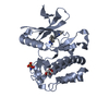 4wnpS S: Starting model for refinement |
|---|---|
| Similar structure data |


-Links
 PDBj
PDBj- Assembly
| Deposited unit | 
| ||||||||
|---|---|---|---|---|---|---|---|---|---|
| 1 |
| ||||||||
| Unit cell |
|
-Components
| #1: Protein | Mass: 31939.977 Da / Num. of mol.: 1 Source method: isolated from a genetically manipulated source Source: (gene. exp.) Homo sapiens (human) / Gene: ULK3 / Production host:   Spodoptera frugiperda (fall armyworm) Spodoptera frugiperda (fall armyworm)References: UniProt: Q6PHR2, non-specific serine/threonine protein kinase | ||
|---|---|---|---|
| #2: Chemical | Bosutinib  Mass: 530.446 Da / Num. of mol.: 2 / Source method: obtained synthetically / Formula: C26H29Cl2N5O3 / Comment: inhibitor, medication*YM Mass: 530.446 Da / Num. of mol.: 2 / Source method: obtained synthetically / Formula: C26H29Cl2N5O3 / Comment: inhibitor, medication*YM#3: Water | ChemComp-HOH / | Water Mass: 18.015 Da / Num. of mol.: 127 / Source method: isolated from a natural source / Formula: H2O Mass: 18.015 Da / Num. of mol.: 127 / Source method: isolated from a natural source / Formula: H2O |
-Experimental details
-Experiment
| Experiment | Method: X-RAY DIFFRACTION / Number of used crystals: 1 |
|---|
- Sample preparation
| Crystal | Density Matthews: 2.81 Å3/Da / Density % sol: 56.26 % |
|---|---|
| Crystal grow | Temperature: 277 K / Method: vapor diffusion, sitting drop / pH: 8.5 Details: 0.1 M Tris pH 8.5; 0.2 M sodium citrate; 30% PEG400 |
-Data collection
| Diffraction | Mean temperature: 100 K |
|---|---|
| Diffraction source | Source: SYNCHROTRON / Site: Diamond  / Beamline: I04-1 / Wavelength: 0.9282 Å / Beamline: I04-1 / Wavelength: 0.9282 Å |
| Detector | Type: DECTRIS PILATUS 6M / Detector: PIXEL / Date: Sep 12, 2016 |
| Radiation | Protocol: SINGLE WAVELENGTH / Monochromatic (M) / Laue (L): M / Scattering type: x-ray |
| Radiation wavelength | Wavelength: 0.9282 Å / Relative weight: 1 |
| Reflection | Resolution: 1.7→48.18 Å / Num. obs: 39968 / % possible obs: 100 % / Redundancy: 6.5 % / Rrim(I) all: 0.09 / Net I/σ(I): 13.4 |
| Reflection shell | Resolution: 1.7→1.73 Å / Redundancy: 6 % / Mean I/σ(I) obs: 2.1 / Num. unique obs: 1951 / Rrim(I) all: 0.824 / % possible all: 99.1 |
- Processing
| Software |
| ||||||||||||||||||||||||||||||||||||||||||||||||||||||||||||||||||||||||||||||||||||||||||||||||||||||||||||||||||||||||||||||||||||||||||||||||||||||||||||||||||||||||||||||||||||||
|---|---|---|---|---|---|---|---|---|---|---|---|---|---|---|---|---|---|---|---|---|---|---|---|---|---|---|---|---|---|---|---|---|---|---|---|---|---|---|---|---|---|---|---|---|---|---|---|---|---|---|---|---|---|---|---|---|---|---|---|---|---|---|---|---|---|---|---|---|---|---|---|---|---|---|---|---|---|---|---|---|---|---|---|---|---|---|---|---|---|---|---|---|---|---|---|---|---|---|---|---|---|---|---|---|---|---|---|---|---|---|---|---|---|---|---|---|---|---|---|---|---|---|---|---|---|---|---|---|---|---|---|---|---|---|---|---|---|---|---|---|---|---|---|---|---|---|---|---|---|---|---|---|---|---|---|---|---|---|---|---|---|---|---|---|---|---|---|---|---|---|---|---|---|---|---|---|---|---|---|---|---|---|---|
| Refinement | Method to determine structure: MOLECULAR REPLACEMENT Starting model: 4wnp Resolution: 1.7→48.18 Å / Cor.coef. Fo:Fc: 0.948 / Cor.coef. Fo:Fc free: 0.938 / SU B: 2.236 / SU ML: 0.073 / Cross valid method: THROUGHOUT / ESU R: 0.099 / ESU R Free: 0.101 / Stereochemistry target values: MAXIMUM LIKELIHOOD / Details: HYDROGENS HAVE BEEN ADDED IN THE RIDING POSITIONS
| ||||||||||||||||||||||||||||||||||||||||||||||||||||||||||||||||||||||||||||||||||||||||||||||||||||||||||||||||||||||||||||||||||||||||||||||||||||||||||||||||||||||||||||||||||||||
| Solvent computation | Ion probe radii: 0.8 Å / Shrinkage radii: 0.8 Å / VDW probe radii: 1.2 Å / Solvent model: MASK | ||||||||||||||||||||||||||||||||||||||||||||||||||||||||||||||||||||||||||||||||||||||||||||||||||||||||||||||||||||||||||||||||||||||||||||||||||||||||||||||||||||||||||||||||||||||
| Displacement parameters | Biso mean: 24.039 Å2
| ||||||||||||||||||||||||||||||||||||||||||||||||||||||||||||||||||||||||||||||||||||||||||||||||||||||||||||||||||||||||||||||||||||||||||||||||||||||||||||||||||||||||||||||||||||||
| Refinement step | Cycle: 1 / Resolution: 1.7→48.18 Å
| ||||||||||||||||||||||||||||||||||||||||||||||||||||||||||||||||||||||||||||||||||||||||||||||||||||||||||||||||||||||||||||||||||||||||||||||||||||||||||||||||||||||||||||||||||||||
| Refine LS restraints |
|