 Movie
Movie Controller

[English] 日本語
 Yorodumi
- PDB-3g51: Structural diversity of the active conformation of the N-terminal... -
+ Open data

- Basic information
| Entry | Database: PDB / ID: 3g51 | ||||||
|---|---|---|---|---|---|---|---|
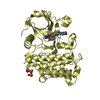
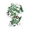
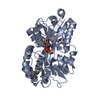
| Title | Structural diversity of the active conformation of the N-terminal kinase domain of p90 ribosomal S6 kinase 2 | ||||||
 Components Components | Ribosomal protein S6 kinase alpha-3 Ribosome Ribosome | ||||||
 Keywords Keywords | TRANSFERASE / N-terminal kinase domain of p90 ribosomal S6 kinase 2 / ATP-binding / Kinase / Nucleotide-binding / Phosphoprotein / Serine/threonine-protein kinase | ||||||
| Function / homology |  Function and homology information Function and homology informationRSK activation / CREB phosphorylation / CREB1 phosphorylation through NMDA receptor-mediated activation of RAS signaling / Gastrin-CREB signalling pathway via PKC and MAPK / Senescence-Associated Secretory Phenotype (SASP) / ribosomal protein S6 kinase activity / ERK/MAPK targets / toll-like receptor signaling pathway / cysteine-type endopeptidase inhibitor activity involved in apoptotic process / response to lipopolysaccharide ...RSK activation / CREB phosphorylation / CREB1 phosphorylation through NMDA receptor-mediated activation of RAS signaling / Gastrin-CREB signalling pathway via PKC and MAPK / Senescence-Associated Secretory Phenotype (SASP) / ribosomal protein S6 kinase activity / ERK/MAPK targets / toll-like receptor signaling pathway / cysteine-type endopeptidase inhibitor activity involved in apoptotic process / response to lipopolysaccharide / non-specific serine/threonine protein kinase / intracellular signal transduction / cell cycle / phosphorylation / protein serine kinase activity / protein serine/threonine kinase activity / nucleolus / protein kinase binding / magnesium ion binding / positive regulation of transcription by RNA polymerase II / nucleoplasm / ATP binding / cytosol / cytoplasmSimilarity search - Function | ||||||
| Biological species |  Mus musculus (house mouse) Mus musculus (house mouse) | ||||||
| Method | X-RAY DIFFRACTION / SYNCHROTRON / MOLECULAR REPLACEMENT / Resolution: 1.8 Å | ||||||
 Authors Authors | Kurinov, I. | ||||||
 Citation Citation | Journal: Plos One / Year: 2009 Title: Structural diversity of the active N-terminal kinase domain of p90 ribosomal S6 kinase 2 Authors: Malakhova, M. / Kurinov, I. / Liu, K. / Zheng, D. / D'Angelo, I. / Shim, J.H. / Steinman, V. / Bode, A.M. / Dong, Z. | ||||||
| History |
|
- Structure visualization
| Structure viewer | Molecule: MolmilJmol/JSmol |
|---|
- Downloads & links
-Download
| PDBx/mmCIF format | 3g51.cif.gz | 78.7 KB | Display | PDBx/mmCIF format |
|---|---|---|---|---|
| PDB format | pdb3g51.ent.gz | 56.8 KB | Display | PDB format |
| PDBx/mmJSON format | 3g51.json.gz | Tree view | PDBx/mmJSON format | |
| Others |  Other downloads Other downloads |
-Validation report
| Arichive directory | https://data.pdbj.org/pub/pdb/validation_reports/g5/3g51ftp://data.pdbj.org/pub/pdb/validation_reports/g5/3g51 | HTTPS FTP |
|---|
-Related structure data
| Related structure data | 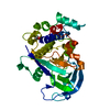 1vzoS S: Starting model for refinement |
|---|---|
| Similar structure data |


-Links
 PDBj
PDBj


- Assembly
| Deposited unit | 
| ||||||||
|---|---|---|---|---|---|---|---|---|---|
| 1 |
| ||||||||
| Unit cell |
|
-Components
| #1: Protein | Ribosome / S6K-alpha 3 / 90 kDa ribosomal protein S6 kinase 3 / p90-RSK 3 / Ribosomal S6 kinase 2 / RSK-2 / ...S6K-alpha 3 / 90 kDa ribosomal protein S6 kinase 3 / p90-RSK 3 / Ribosomal S6 kinase 2 / RSK-2 / pp90RSK2 / MAP kinase-activated protein kinase 1b / MAPKAPK1B Mass: 37475.418 Da / Num. of mol.: 1 Source method: isolated from a genetically manipulated source Source: (gene. exp.) Mus musculus (house mouse)Gene: Mus musculus ribosomal protein S6 kinase 2, Rps6ka-rs1, Rps6ka3, Rsk2 Production host:  Escherichia coli (E. coli) / Strain (production host): BL21-Codon Plus (DE3)-RILP Escherichia coli (E. coli) / Strain (production host): BL21-Codon Plus (DE3)-RILPReferences: UniProt: P18654, non-specific serine/threonine protein kinase |
|---|---|
| #2: Chemical | ChemComp-ANP /   Mass: 506.196 Da / Num. of mol.: 1 / Source method: obtained synthetically / Formula: C10H17N6O12P3 / Comment: AMP-PNP, energy-carrying molecule analogue*YM Mass: 506.196 Da / Num. of mol.: 1 / Source method: obtained synthetically / Formula: C10H17N6O12P3 / Comment: AMP-PNP, energy-carrying molecule analogue*YM |
| #3: Water | ChemComp-HOH / Water Mass: 18.015 Da / Num. of mol.: 274 / Source method: isolated from a natural source / Formula: H2O Mass: 18.015 Da / Num. of mol.: 274 / Source method: isolated from a natural source / Formula: H2O |
-Experimental details
-Experiment
| Experiment | Method: X-RAY DIFFRACTION / Number of used crystals: 1 |
|---|
- Sample preparation
| Crystal | Density Matthews: 2.47 Å3/Da / Density % sol: 50.22 % |
|---|---|
| Crystal grow | Temperature: 293 K / Method: vapor diffusion, sitting drop / pH: 7.5 Details: The diluted protein at different concentrations (2.5 mg/ml 10 mg/ml) was mixed at a 2:1 ratio with precipitant solution (6%-10% PEG 3350, 0.2 M proline, 0.1 M Hepes pH 7.5). , VAPOR ...Details: The diluted protein at different concentrations (2.5 mg/ml 10 mg/ml) was mixed at a 2:1 ratio with precipitant solution (6%-10% PEG 3350, 0.2 M proline, 0.1 M Hepes pH 7.5). , VAPOR DIFFUSION, SITTING DROP, temperature 293K |
-Data collection
| Diffraction | Mean temperature: 100 K |
|---|---|
| Diffraction source | Source: SYNCHROTRON / Site: APS  / Beamline: 24-ID-E / Wavelength: 0.979 Å / Beamline: 24-ID-E / Wavelength: 0.979 Å |
| Detector | Type: ADSC QUANTUM 315 / Detector: CCD / Date: Mar 7, 2008 / Details: HFM, VFM, microdiffractometer |
| Radiation | Monochromator: Si 111 Double Crystal Monochromator / Protocol: SINGLE WAVELENGTH / Monochromatic (M) / Laue (L): M / Scattering type: x-ray |
| Radiation wavelength | Wavelength: 0.979 Å / Relative weight: 1 |
| Reflection | Resolution: 1.8→36 Å / Num. all: 35155 / Num. obs: 35092 / % possible obs: 99.4 % / Observed criterion σ(F): 0 / Observed criterion σ(I): 0 / Redundancy: 6.2 % / Rmerge(I) obs: 0.091 |
| Reflection shell | Resolution: 1.8→1.86 Å / Redundancy: 5 % / % possible all: 94.8 |
- Processing
| Software |
| |||||||||||||||||||||||||
|---|---|---|---|---|---|---|---|---|---|---|---|---|---|---|---|---|---|---|---|---|---|---|---|---|---|---|
| Refinement | Method to determine structure: MOLECULAR REPLACEMENT Starting model: PDB entry 1VZO Resolution: 1.8→36.3 Å / Isotropic thermal model: Isotropic / Cross valid method: THROUGHOUT / σ(F): 0 / σ(I): 0 / Stereochemistry target values: Engh & Huber
| |||||||||||||||||||||||||
| Refinement step | Cycle: LAST / Resolution: 1.8→36.3 Å
| |||||||||||||||||||||||||
| Refine LS restraints |
|
