 Movie
Movie Controller


+ Open data

- Basic information
| Entry | Database: PDB / ID: 6f37 | ||||||
|---|---|---|---|---|---|---|---|
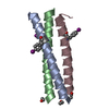
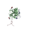
| Title | Fusion protein of RSL and trimeric coiled coil | ||||||
 Components Components | Nano3,Fucose-binding lectin protein | ||||||
 Keywords Keywords | SUGAR BINDING PROTEIN / genetic fusion /  coiled-coil / lectin / trimer coiled-coil / lectin / trimer | ||||||
| Function / homology | Fucose-specific lectin / Fungal fucose-specific lectin / carbohydrate binding / beta-D-mannopyranose / Fucose-binding lectin protein Function and homology information Function and homology information | ||||||
| Biological species |  Ralstonia solanacearum (bacteria) Ralstonia solanacearum (bacteria) | ||||||
| Method | X-RAY DIFFRACTION / SYNCHROTRON / MOLECULAR REPLACEMENT / molecular replacement / Resolution: 2.2 Å | ||||||
 Authors Authors | Wronska, M.A. / Rennie, M.L. / Crowley, P.B. | ||||||
| Funding support |  Ireland, 1items Ireland, 1items
| ||||||
 Citation Citation | Journal: To Be Published Title: Fusion protein of RSL and trimeric coiled coil Authors: Wronska, M.A. / Rennie, M.L. / Crowley, P.B. | ||||||
| History |
|
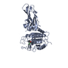
- Structure visualization
| Structure viewer | Molecule: MolmilJmol/JSmol |
|---|
- Downloads & links
-Download
| PDBx/mmCIF format | 6f37.cif.gz | 59.5 KB | Display | PDBx/mmCIF format |
|---|---|---|---|---|
| PDB format | pdb6f37.ent.gz | 41.9 KB | Display | PDB format |
| PDBx/mmJSON format | 6f37.json.gz | Tree view | PDBx/mmJSON format | |
| Others |  Other downloads Other downloads |
-Validation report
| Arichive directory | https://data.pdbj.org/pub/pdb/validation_reports/f3/6f37ftp://data.pdbj.org/pub/pdb/validation_reports/f3/6f37 | HTTPS FTP |
|---|
-Related structure data
| Related structure data | 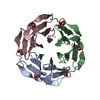 2bt9S S: Starting model for refinement |
|---|---|
| Similar structure data |


-Links
 PDBj
PDBj- Assembly
| Deposited unit | 
| ||||||||
|---|---|---|---|---|---|---|---|---|---|
| 1 | 
| ||||||||
| 2 | 
| ||||||||
| Unit cell |
| ||||||||
| Components on special symmetry positions |
|
-Components
| #1: Protein | Mass: 12157.452 Da / Num. of mol.: 2 Source method: isolated from a genetically manipulated source Source: (gene. exp.) Ralstonia solanacearum (bacteria) / Plasmid: pET-25b(+) / Gene: RSP795_21825, RSP799_05830, RUN39_v1_50103 / Production host: Escherichia coli BL21(DE3) (bacteria) / References: UniProt: A0A0S4TLR1#2: Sugar | ChemComp-BMA / Mannose  Type: D-saccharide, beta linking / Mass: 180.156 Da / Num. of mol.: 4 Type: D-saccharide, beta linking / Mass: 180.156 Da / Num. of mol.: 4Source method: isolated from a genetically manipulated source Formula: C6H12O6 #3: Chemical | ChemComp-NHE / | CHES (buffer)  Mass: 207.290 Da / Num. of mol.: 1 / Source method: obtained synthetically / Formula: C8H17NO3S / Comment: pH buffer*YM Mass: 207.290 Da / Num. of mol.: 1 / Source method: obtained synthetically / Formula: C8H17NO3S / Comment: pH buffer*YM#4: Water | ChemComp-HOH / | Water Mass: 18.015 Da / Num. of mol.: 74 / Source method: isolated from a natural source / Formula: H2O Mass: 18.015 Da / Num. of mol.: 74 / Source method: isolated from a natural source / Formula: H2O |
|---|
-Experimental details
-Experiment
| Experiment | Method: X-RAY DIFFRACTION / Number of used crystals: 1 |
|---|
- Sample preparation
| Crystal | Density Matthews: 2.4 Å3/Da / Density % sol: 48.68 % / Description: Cubic |
|---|---|
| Crystal grow | Temperature: 293.15 K / Method: vapor diffusion, sitting drop / pH: 9.5 / Details: 20% PEG 8000, 0.1 M CHES pH 9.5 |
-Data collection
| Diffraction | Mean temperature: 100 K | ||||||||||||||||||||||||
|---|---|---|---|---|---|---|---|---|---|---|---|---|---|---|---|---|---|---|---|---|---|---|---|---|---|
| Diffraction source | Source: SYNCHROTRON / Site: SOLEIL  / Beamline: PROXIMA 2 / Wavelength: 0.98 Å / Beamline: PROXIMA 2 / Wavelength: 0.98 Å | ||||||||||||||||||||||||
| Detector | Type: DECTRIS EIGER X 9M / Detector: PIXEL / Date: Feb 11, 2017 | ||||||||||||||||||||||||
| Radiation | Protocol: SINGLE WAVELENGTH / Monochromatic (M) / Laue (L): M / Scattering type: x-ray | ||||||||||||||||||||||||
| Radiation wavelength | Wavelength: 0.98 Å / Relative weight: 1 | ||||||||||||||||||||||||
| Reflection | Resolution: 2.2→88.76 Å / Num. obs: 12123 / % possible obs: 100 % / Redundancy: 13.2 % / CC1/2: 0.997 / Rmerge(I) obs: 0.178 / Rpim(I) all: 0.051 / Rrim(I) all: 0.185 / Net I/σ(I): 21.6 | ||||||||||||||||||||||||
| Reflection shell | Diffraction-ID: 1
|
-Phasing
| Phasing | Method: molecular replacement |
|---|
- Processing
| Software |
| ||||||||||||||||||||||||||||||||||||||||||||||||||||||||||||
|---|---|---|---|---|---|---|---|---|---|---|---|---|---|---|---|---|---|---|---|---|---|---|---|---|---|---|---|---|---|---|---|---|---|---|---|---|---|---|---|---|---|---|---|---|---|---|---|---|---|---|---|---|---|---|---|---|---|---|---|---|---|
| Refinement | Method to determine structure: MOLECULAR REPLACEMENT Starting model: 2BT9 chain A Resolution: 2.2→62.81 Å / Cor.coef. Fo:Fc: 0.948 / Cor.coef. Fo:Fc free: 0.928 / SU B: 7.352 / SU ML: 0.178 / SU R Cruickshank DPI: 0.3069 / Cross valid method: THROUGHOUT / σ(F): 0 / ESU R: 0.307 / ESU R Free: 0.22 Details: HYDROGENS HAVE BEEN ADDED IN THE RIDING POSITIONS U VALUES : REFINED INDIVIDUALLY
| ||||||||||||||||||||||||||||||||||||||||||||||||||||||||||||
| Solvent computation | Ion probe radii: 0.8 Å / Shrinkage radii: 0.8 Å / VDW probe radii: 1.2 Å | ||||||||||||||||||||||||||||||||||||||||||||||||||||||||||||
| Displacement parameters | Biso max: 291.94 Å2 / Biso mean: 61.227 Å2 / Biso min: 20.12 Å2
| ||||||||||||||||||||||||||||||||||||||||||||||||||||||||||||
| Refinement step | Cycle: final / Resolution: 2.2→62.81 Å
| ||||||||||||||||||||||||||||||||||||||||||||||||||||||||||||
| Refine LS restraints |
| ||||||||||||||||||||||||||||||||||||||||||||||||||||||||||||
| LS refinement shell | Resolution: 2.201→2.258 Å / Rfactor Rfree error: 0 / Total num. of bins used: 20
|
