 Movie
Movie Controller


+ Open data

- Basic information
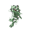
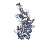
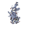
| Entry | Database: PDB / ID: 1q3x | ||||||
|---|---|---|---|---|---|---|---|
| Title | Crystal structure of the catalytic region of human MASP-2 | ||||||
 Components Components | Mannan-binding lectin serine protease 2 | ||||||
 Keywords Keywords |  HYDROLASE / complement / serine protease / modular structure / hinge bending / autoactivation HYDROLASE / complement / serine protease / modular structure / hinge bending / autoactivation | ||||||
| Function / homology |  Function and homology informationmannan-binding lectin-associated serine protease-2 / complement component C4b binding / Ficolins bind to repetitive carbohydrate structures on the target cell surface / Lectin pathway of complement activation / complement activation, lectin pathway / Initial triggering of complement / complement activation, classical pathway / calcium-dependent protein binding / peptidase activity / serine-type endopeptidase activity ...mannan-binding lectin-associated serine protease-2 / complement component C4b binding / Ficolins bind to repetitive carbohydrate structures on the target cell surface / Lectin pathway of complement activation / complement activation, lectin pathway / Initial triggering of complement / complement activation, classical pathway / calcium-dependent protein binding / peptidase activity / serine-type endopeptidase activity / calcium ion binding / SARS-CoV-2 activates/modulates innate and adaptive immune responses / proteolysis / extracellular space / extracellular exosome / extracellular region / identical protein binding Function and homology informationmannan-binding lectin-associated serine protease-2 / complement component C4b binding / Ficolins bind to repetitive carbohydrate structures on the target cell surface / Lectin pathway of complement activation / complement activation, lectin pathway / Initial triggering of complement / complement activation, classical pathway / calcium-dependent protein binding / peptidase activity / serine-type endopeptidase activity ...mannan-binding lectin-associated serine protease-2 / complement component C4b binding / Ficolins bind to repetitive carbohydrate structures on the target cell surface / Lectin pathway of complement activation / complement activation, lectin pathway / Initial triggering of complement / complement activation, classical pathway / calcium-dependent protein binding / peptidase activity / serine-type endopeptidase activity / calcium ion binding / SARS-CoV-2 activates/modulates innate and adaptive immune responses / proteolysis / extracellular space / extracellular exosome / extracellular region / identical protein bindingSimilarity search - Function | ||||||
| Biological species |  Homo sapiens (human) Homo sapiens (human) | ||||||
| Method | X-RAY DIFFRACTION / SYNCHROTRON / MOLECULAR REPLACEMENT / Resolution: 2.23 Å | ||||||
 Authors Authors | Harmat, V. / Gal, P. / Kardos, J. / Szilagyi, K. / Ambrus, G. / Naray-Szabo, G. / Zavodszky, P. | ||||||
 Citation Citation | Journal: J.Mol.Biol. / Year: 2004 Title: The structure of MBL-associated serine protease-2 reveals that identical substrate specificities of C1s and MASP-2 are realized through different sets of enzyme-substrate interactions Authors: Harmat, V. / Gal, P. / Kardos, J. / Szilagyi, K. / Ambrus, G. / Vegh, B. / Naray-Szabo, G. / Zavodsky, P. #1: Journal: J.Immunol. / Year: 2003Title: Natural substrates and inhibitors of mannan-binding lectin-associated serine protease 1 and 2: A study on recombinant catalytic fragments Authors: Ambrus, G. / Gal, P. / Kojima, M. / Szilagyi, K. / Balczer, J. / Antal, J. / Graf, L. / Laich, A. / Moffat, B.E. / Schwaelbe, W. / Sim, R.B. / Zvodszky, P. #2: Journal: Embo J. / Year: 2000Title: Crystal structure of the catalytic domain of human complement C1s: a serine protease with a handle Authors: Gaboriaud, C. / Rossi, V. / Bally, I. / Arlaud, G.J. / Fontecilla-Camps, J.C. | ||||||
| History |
|
- Structure visualization
| Structure viewer | Molecule: MolmilJmol/JSmol |
|---|
- Downloads & links
-Download
| PDBx/mmCIF format | 1q3x.cif.gz | 146.3 KB | Display | PDBx/mmCIF format |
|---|---|---|---|---|
| PDB format | pdb1q3x.ent.gz | 112.4 KB | Display | PDB format |
| PDBx/mmJSON format | 1q3x.json.gz | Tree view | PDBx/mmJSON format | |
| Others |  Other downloads Other downloads |
-Validation report
| Arichive directory | https://data.pdbj.org/pub/pdb/validation_reports/q3/1q3xftp://data.pdbj.org/pub/pdb/validation_reports/q3/1q3x | HTTPS FTP |
|---|
-Related structure data
| Related structure data |  1elvS S: Starting model for refinement |
|---|---|
| Similar structure data |


-Links
 PDBj
PDBj


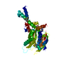
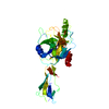
- Assembly
| Deposited unit | 
| |||||||||||||||||||||||||||||||||||||||||||||||||||||||||||||||||||||||||||||||||||||||||||||||||||||||||||||||||||||||||||||||||||||||||||||||||||||||||||||||||||||||||||||||||||||||||||||
|---|---|---|---|---|---|---|---|---|---|---|---|---|---|---|---|---|---|---|---|---|---|---|---|---|---|---|---|---|---|---|---|---|---|---|---|---|---|---|---|---|---|---|---|---|---|---|---|---|---|---|---|---|---|---|---|---|---|---|---|---|---|---|---|---|---|---|---|---|---|---|---|---|---|---|---|---|---|---|---|---|---|---|---|---|---|---|---|---|---|---|---|---|---|---|---|---|---|---|---|---|---|---|---|---|---|---|---|---|---|---|---|---|---|---|---|---|---|---|---|---|---|---|---|---|---|---|---|---|---|---|---|---|---|---|---|---|---|---|---|---|---|---|---|---|---|---|---|---|---|---|---|---|---|---|---|---|---|---|---|---|---|---|---|---|---|---|---|---|---|---|---|---|---|---|---|---|---|---|---|---|---|---|---|---|---|---|---|---|---|---|
| 1 | 
| |||||||||||||||||||||||||||||||||||||||||||||||||||||||||||||||||||||||||||||||||||||||||||||||||||||||||||||||||||||||||||||||||||||||||||||||||||||||||||||||||||||||||||||||||||||||||||||
| 2 | 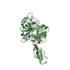
| |||||||||||||||||||||||||||||||||||||||||||||||||||||||||||||||||||||||||||||||||||||||||||||||||||||||||||||||||||||||||||||||||||||||||||||||||||||||||||||||||||||||||||||||||||||||||||||
| Unit cell |
| |||||||||||||||||||||||||||||||||||||||||||||||||||||||||||||||||||||||||||||||||||||||||||||||||||||||||||||||||||||||||||||||||||||||||||||||||||||||||||||||||||||||||||||||||||||||||||||
| Noncrystallographic symmetry (NCS) | NCS domain:
NCS domain segments: Ens-ID: 1 / Refine code: 1
|