 Movie
Movie Controller


+ Open data

- Basic information
| Entry | Database: PDB / ID: 6es2 | ||||||
|---|---|---|---|---|---|---|---|
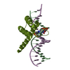
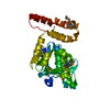
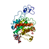
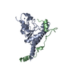
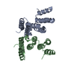
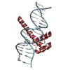
| Title | Structure of CDX2-DNA(CAA) | ||||||
 Components Components |
| ||||||
 Keywords Keywords |  TRANSCRIPTION / homeodomain transcription factor / CDX2-DNA complex TRANSCRIPTION / homeodomain transcription factor / CDX2-DNA complex | ||||||
| Function / homology |  Function and homology information Function and homology informationintestinal epithelial cell differentiation / regulation of somitogenesis / trophectodermal cell differentiation / labyrinthine layer development / establishment or maintenance of epithelial cell apical/basal polarity / methyl-CpG binding / anterior/posterior axis specification / endosome to lysosome transport / blood vessel development / somatic stem cell population maintenance ...intestinal epithelial cell differentiation / regulation of somitogenesis / trophectodermal cell differentiation / labyrinthine layer development / establishment or maintenance of epithelial cell apical/basal polarity / methyl-CpG binding / anterior/posterior axis specification / endosome to lysosome transport / blood vessel development / somatic stem cell population maintenance / transcription repressor complex / condensed nuclear chromosome / stem cell differentiation / positive regulation of cell differentiation / RNA polymerase II transcription regulatory region sequence-specific DNA binding / animal organ morphogenesis / Synthesis, secretion, and inactivation of Glucagon-like Peptide-1 (GLP-1) / DNA-binding transcription repressor activity, RNA polymerase II-specific / sequence-specific double-stranded DNA binding / cell differentiation / DNA-binding transcription factor activity, RNA polymerase II-specific / RNA polymerase II cis-regulatory region sequence-specific DNA binding / DNA-binding transcription factor activity / positive regulation of cell population proliferation / chromatin / regulation of transcription by RNA polymerase II / negative regulation of transcription by RNA polymerase II / positive regulation of transcription by RNA polymerase II / nucleoplasm / nucleusSimilarity search - Function | ||||||
| Biological species |  Homo sapiens (human) Homo sapiens (human) | ||||||
| Method | X-RAY DIFFRACTION / SYNCHROTRON / MOLECULAR REPLACEMENT / Resolution: 2.95 Å | ||||||
 Authors Authors | Morgunova, E. / Yin, Y. / Jolma, A. / Popov, A. / Taipale, J. | ||||||
 Citation Citation | Journal: Elife / Year: 2018 Title: Two distinct DNA sequences recognized by transcription factors represent enthalpy and entropy optima. Authors: Morgunova, E. / Yin, Y. / Das, P.K. / Jolma, A. / Zhu, F. / Popov, A. / Xu, Y. / Nilsson, L. / Taipale, J. | ||||||
| History |
|
- Structure visualization
| Structure viewer | Molecule: MolmilJmol/JSmol |
|---|
- Downloads & links
-Download
| PDBx/mmCIF format | 6es2.cif.gz | 154.8 KB | Display | PDBx/mmCIF format |
|---|---|---|---|---|
| PDB format | pdb6es2.ent.gz | 120.4 KB | Display | PDB format |
| PDBx/mmJSON format | 6es2.json.gz | Tree view | PDBx/mmJSON format | |
| Others |  Other downloads Other downloads |
-Validation report
| Arichive directory | https://data.pdbj.org/pub/pdb/validation_reports/es/6es2ftp://data.pdbj.org/pub/pdb/validation_reports/es/6es2 | HTTPS FTP |
|---|
-Related structure data
| Related structure data |  5ednC 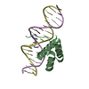 5eeaC  6es3C  5ltyS S: Starting model for refinement C: citing same article ( |
|---|---|
| Similar structure data |

-Links
 PDBj
PDBj


- Assembly
| Deposited unit | 
| ||||||||||||||||||||||||||||||||||||||||||||||||
|---|---|---|---|---|---|---|---|---|---|---|---|---|---|---|---|---|---|---|---|---|---|---|---|---|---|---|---|---|---|---|---|---|---|---|---|---|---|---|---|---|---|---|---|---|---|---|---|---|---|
| 1 | 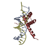
| ||||||||||||||||||||||||||||||||||||||||||||||||
| 2 | 
| ||||||||||||||||||||||||||||||||||||||||||||||||
| Unit cell |
| ||||||||||||||||||||||||||||||||||||||||||||||||
| Noncrystallographic symmetry (NCS) | NCS domain:
NCS domain segments:
NCS ensembles :
|
-Components
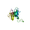
| #1: DNA chain | Mass: 5454.521 Da / Num. of mol.: 2 / Source method: obtained synthetically / Source: (synth.) Homo sapiens (human)#2: Protein | / CDX-3 / Caudal-type homeobox protein 2Mass: 8896.251 Da / Num. of mol.: 2 Source method: isolated from a genetically manipulated source Source: (gene. exp.) Homo sapiens (human) / Gene: CDX2, CDX3 / Plasmid: pETG20A / Production host:  Escherichia coli (E. coli) / References: UniProt: Q99626 Escherichia coli (E. coli) / References: UniProt: Q99626#3: DNA chain | Mass: 5575.672 Da / Num. of mol.: 2 / Source method: obtained synthetically / Source: (synth.) Homo sapiens (human)#4: Water | ChemComp-HOH / | Water Mass: 18.015 Da / Num. of mol.: 10 / Source method: isolated from a natural source / Formula: H2O Mass: 18.015 Da / Num. of mol.: 10 / Source method: isolated from a natural source / Formula: H2O |
|---|
-Experimental details
-Experiment
| Experiment | Method: X-RAY DIFFRACTION / Number of used crystals: 1 |
|---|
- Sample preparation
| Crystal | Density Matthews: 2.61 Å3/Da / Density % sol: 52.92 % |
|---|---|
| Crystal grow | Temperature: 298 K / Method: vapor diffusion, sitting drop / pH: 8 Details: 27 % PME 5000, 0.15M potassium chloride, 0.1M magnesium chloride, 8% PEG 400, 0.05M TRIS buffer |
-Data collection
| Diffraction | Mean temperature: 100 K | ||||||||||||||||||||||||
|---|---|---|---|---|---|---|---|---|---|---|---|---|---|---|---|---|---|---|---|---|---|---|---|---|---|
| Diffraction source | Source: SYNCHROTRON / Site: ESRF  / Beamline: ID23-1 / Wavelength: 0.97242 Å / Beamline: ID23-1 / Wavelength: 0.97242 Å | ||||||||||||||||||||||||
| Detector | Type: DECTRIS PILATUS 6M-F / Detector: PIXEL / Date: Jul 2, 2016 | ||||||||||||||||||||||||
| Radiation | Protocol: SINGLE WAVELENGTH / Monochromatic (M) / Laue (L): M / Scattering type: x-ray | ||||||||||||||||||||||||
| Radiation wavelength | Wavelength: 0.97242 Å / Relative weight: 1 | ||||||||||||||||||||||||
| Reflection | Resolution: 2.95→66.19 Å / Num. obs: 8494 / % possible obs: 96.6 % / Redundancy: 3.2 % / CC1/2: 0.998 / Rmerge(I) obs: 0.077 / Rpim(I) all: 0.051 / Rrim(I) all: 0.093 / Net I/σ(I): 7.5 | ||||||||||||||||||||||||
| Reflection shell | Diffraction-ID: 1
|
- Processing
| Software |
| |||||||||||||||||||||||||||||||||||||||||||||||||||||||||||||||||||||||||||||||||||||||||||||||||||||||||||||||||||||||||||||
|---|---|---|---|---|---|---|---|---|---|---|---|---|---|---|---|---|---|---|---|---|---|---|---|---|---|---|---|---|---|---|---|---|---|---|---|---|---|---|---|---|---|---|---|---|---|---|---|---|---|---|---|---|---|---|---|---|---|---|---|---|---|---|---|---|---|---|---|---|---|---|---|---|---|---|---|---|---|---|---|---|---|---|---|---|---|---|---|---|---|---|---|---|---|---|---|---|---|---|---|---|---|---|---|---|---|---|---|---|---|---|---|---|---|---|---|---|---|---|---|---|---|---|---|---|---|---|
| Refinement | Method to determine structure: MOLECULAR REPLACEMENT Starting model: 5LTY Resolution: 2.95→66.19 Å / Cor.coef. Fo:Fc: 0.964 / Cor.coef. Fo:Fc free: 0.948 / SU B: 53.863 / SU ML: 0.427 / Cross valid method: THROUGHOUT / σ(F): 0 / ESU R Free: 0.431 Details: HYDROGENS HAVE BEEN ADDED IN THE RIDING POSITIONS U VALUES : WITH TLS ADDED
| |||||||||||||||||||||||||||||||||||||||||||||||||||||||||||||||||||||||||||||||||||||||||||||||||||||||||||||||||||||||||||||
| Solvent computation | Ion probe radii: 0.8 Å / Shrinkage radii: 0.8 Å / VDW probe radii: 1.2 Å | |||||||||||||||||||||||||||||||||||||||||||||||||||||||||||||||||||||||||||||||||||||||||||||||||||||||||||||||||||||||||||||
| Displacement parameters | Biso max: 204.74 Å2 / Biso mean: 98.684 Å2 / Biso min: 57.54 Å2
| |||||||||||||||||||||||||||||||||||||||||||||||||||||||||||||||||||||||||||||||||||||||||||||||||||||||||||||||||||||||||||||
| Refinement step | Cycle: final / Resolution: 2.95→66.19 Å
| |||||||||||||||||||||||||||||||||||||||||||||||||||||||||||||||||||||||||||||||||||||||||||||||||||||||||||||||||||||||||||||
| Refine LS restraints |
| |||||||||||||||||||||||||||||||||||||||||||||||||||||||||||||||||||||||||||||||||||||||||||||||||||||||||||||||||||||||||||||
| Refine LS restraints NCS | Refine-ID: X-RAY DIFFRACTION / Type: interatomic distance / Rms dev position: 0.06 Å / Weight position: 0.05
| |||||||||||||||||||||||||||||||||||||||||||||||||||||||||||||||||||||||||||||||||||||||||||||||||||||||||||||||||||||||||||||
| LS refinement shell | Resolution: 2.953→3.029 Å / Rfactor Rfree error: 0 / Total num. of bins used: 20
| |||||||||||||||||||||||||||||||||||||||||||||||||||||||||||||||||||||||||||||||||||||||||||||||||||||||||||||||||||||||||||||
| Refinement TLS params. | Method: refined / Refine-ID: X-RAY DIFFRACTION
| |||||||||||||||||||||||||||||||||||||||||||||||||||||||||||||||||||||||||||||||||||||||||||||||||||||||||||||||||||||||||||||
| Refinement TLS group |
|
