 Movie
Movie Controller

[English] 日本語
 Yorodumi
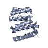
- PDB-4h7x: Crystal structure of the tetratricopeptide repeat (TPR) motif of ... -
+ Open data

- Basic information
| Entry | Database: PDB / ID: 4h7x | ||||||
|---|---|---|---|---|---|---|---|
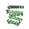
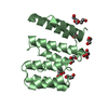
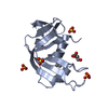
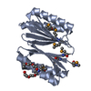
| Title | Crystal structure of the tetratricopeptide repeat (TPR) motif of human dual specificity protein kinase Mps1 | ||||||
 Components Components | Dual specificity protein kinase TTK | ||||||
 Keywords Keywords |  TRANSFERASE / mitotic checkpoint kinase / chromosome instability / cancer / tetratricopeptide repeat (TPR) motif / mitotic checkpoint TRANSFERASE / mitotic checkpoint kinase / chromosome instability / cancer / tetratricopeptide repeat (TPR) motif / mitotic checkpoint | ||||||
| Function / homology |  Function and homology information Function and homology informationprotein localization to meiotic spindle midzone / meiotic spindle assembly checkpoint signaling / kinetochore binding / female meiosis chromosome segregation / protein localization to kinetochore / dual-specificity kinase / spindle organization / mitotic spindle assembly checkpoint signaling / protein serine/threonine/tyrosine kinase activity / mitotic spindle organization ...protein localization to meiotic spindle midzone / meiotic spindle assembly checkpoint signaling / kinetochore binding / female meiosis chromosome segregation / protein localization to kinetochore / dual-specificity kinase / spindle organization / mitotic spindle assembly checkpoint signaling / protein serine/threonine/tyrosine kinase activity / mitotic spindle organization / chromosome segregation / kinetochore / spindle / protein tyrosine kinase activity / phosphorylation / protein serine kinase activity / protein serine/threonine kinase activity / positive regulation of cell population proliferation / ATP binding / membrane / identical protein binding / nucleus / cytoplasmSimilarity search - Function | ||||||
| Biological species |  Homo sapiens (human) Homo sapiens (human) | ||||||
| Method | X-RAY DIFFRACTION / SYNCHROTRON / SAD / Resolution: 2.6 Å | ||||||
 Authors Authors | Bolanos-Garcia, V.M. / Chirgadze, D.Y. / Blundell, T.L. | ||||||
 Citation Citation | Journal: Biochem.J. / Year: 2012 Title: Structural and functional insights into the role of the N-terminal Mps1 TPR domain in the SAC (spindle assembly checkpoint). Authors: Thebault, P. / Chirgadze, D.Y. / Dou, Z. / Blundell, T.L. / Elowe, S. / Bolanos-Garcia, V.M. | ||||||
| History |
|
- Structure visualization
| Structure viewer | Molecule: MolmilJmol/JSmol |
|---|
- Downloads & links
-Download
| PDBx/mmCIF format | 4h7x.cif.gz | 66.4 KB | Display | PDBx/mmCIF format |
|---|---|---|---|---|
| PDB format | pdb4h7x.ent.gz | 50 KB | Display | PDB format |
| PDBx/mmJSON format | 4h7x.json.gz | Tree view | PDBx/mmJSON format | |
| Others |  Other downloads Other downloads |
-Validation report
| Arichive directory | https://data.pdbj.org/pub/pdb/validation_reports/h7/4h7xftp://data.pdbj.org/pub/pdb/validation_reports/h7/4h7x | HTTPS FTP |
|---|
-Related structure data


-Links
 PDBj
PDBj


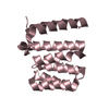
- Assembly
| Deposited unit | 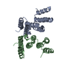
| ||||||||
|---|---|---|---|---|---|---|---|---|---|
| 1 | 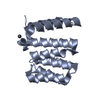
| ||||||||
| 2 | 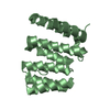
| ||||||||
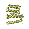
| Unit cell |
|
-Components
| #1: Protein | Mass: 18187.055 Da / Num. of mol.: 2 / Fragment: TPR domain (UNP residues 55-210) Source method: isolated from a genetically manipulated source Source: (gene. exp.) Homo sapiens (human)Gene: MPS1, MPS1L1, NCBI Reference Sequence NM_003318.4, TTK Plasmid: pGST-TPR-Mps1 / Production host:  Escherichia coli (E. coli) / Strain (production host): BL21(DE3) / References: UniProt: P33981, dual-specificity kinase Escherichia coli (E. coli) / Strain (production host): BL21(DE3) / References: UniProt: P33981, dual-specificity kinase#2: Chemical | ChemComp-PB / | Lead  Mass: 207.200 Da / Num. of mol.: 1 / Source method: obtained synthetically / Formula: Pb Mass: 207.200 Da / Num. of mol.: 1 / Source method: obtained synthetically / Formula: Pb#3: Water | ChemComp-HOH / | Water Mass: 18.015 Da / Num. of mol.: 52 / Source method: isolated from a natural source / Formula: H2O Mass: 18.015 Da / Num. of mol.: 52 / Source method: isolated from a natural source / Formula: H2O |
|---|
-Experimental details
-Experiment
| Experiment | Method: X-RAY DIFFRACTION / Number of used crystals: 1 |
|---|
- Sample preparation
| Crystal | Density Matthews: 2.99 Å3/Da / Density % sol: 58.86 % |
|---|---|
| Crystal grow | Temperature: 291 K / Method: vapor diffusion / pH: 6 Details: 20% PEG 3350, 100 mM MES, 250 mM KCl, pH 6.0, VAPOR DIFFUSION, temperature 291K |
-Data collection
| Diffraction | Mean temperature: 100 K |
|---|---|
| Diffraction source | Source: SYNCHROTRON / Site: Diamond  / Beamline: I04 / Wavelength: 0.9497 Å / Beamline: I04 / Wavelength: 0.9497 Å |
| Detector | Type: ADSC QUANTUM 315 / Detector: CCD / Date: Feb 1, 2012 |
| Radiation | Protocol: SINGLE WAVELENGTH / Monochromatic (M) / Laue (L): M / Scattering type: x-ray |
| Radiation wavelength | Wavelength: 0.9497 Å / Relative weight: 1 |
| Reflection | Resolution: 2.6→39.7 Å / Num. all: 14021 / Num. obs: 14021 / % possible obs: 100 % / Observed criterion σ(F): 0 / Observed criterion σ(I): 0 / Redundancy: 10.7 % / Biso Wilson estimate: 35.9 Å2 / Rmerge(I) obs: 0.156 / Rsym value: 0.156 / Net I/σ(I): 7.2 |
| Reflection shell | Resolution: 2.6→2.66 Å / Redundancy: 5.1 % / Rmerge(I) obs: 0.599 / % possible all: 99.9 |
- Processing
| Software |
| |||||||||||||||||||||||||||||||||||||||||||||||||||||||||||||||||||||||||||||
|---|---|---|---|---|---|---|---|---|---|---|---|---|---|---|---|---|---|---|---|---|---|---|---|---|---|---|---|---|---|---|---|---|---|---|---|---|---|---|---|---|---|---|---|---|---|---|---|---|---|---|---|---|---|---|---|---|---|---|---|---|---|---|---|---|---|---|---|---|---|---|---|---|---|---|---|---|---|---|
| Refinement | Method to determine structure: SAD / Resolution: 2.6→39.7 Å / SU ML: 0.29 / σ(F): 0 / Phase error: 21.72 / Stereochemistry target values: MLHL
| |||||||||||||||||||||||||||||||||||||||||||||||||||||||||||||||||||||||||||||
| Solvent computation | Shrinkage radii: 0.73 Å / VDW probe radii: 1 Å / Solvent model: FLAT BULK SOLVENT MODEL / Bsol: 32.41 Å2 / ksol: 0.365 e/Å3 | |||||||||||||||||||||||||||||||||||||||||||||||||||||||||||||||||||||||||||||
| Displacement parameters |
| |||||||||||||||||||||||||||||||||||||||||||||||||||||||||||||||||||||||||||||
| Refinement step | Cycle: LAST / Resolution: 2.6→39.7 Å
| |||||||||||||||||||||||||||||||||||||||||||||||||||||||||||||||||||||||||||||
| Refine LS restraints |
| |||||||||||||||||||||||||||||||||||||||||||||||||||||||||||||||||||||||||||||
| LS refinement shell |
|
