 Movie
Movie Controller

[English] 日本語
 Yorodumi
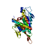
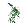
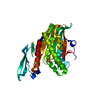
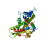
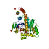
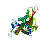
- PDB-6lnr: Structure of intact chitinase with hevein domain from the plant S... -
+ Open data

- Basic information
| Entry | Database: PDB / ID: 6lnr | ||||||
|---|---|---|---|---|---|---|---|
| Title | Structure of intact chitinase with hevein domain from the plant Simarouba glauca, known for its traditional anti-inflammatory efficacy | ||||||
 Components Components | Class-1 chitinase | ||||||
 Keywords Keywords |  HYDROLASE / Chitinase / Hevein HYDROLASE / Chitinase / Hevein | ||||||
| Biological species |  Simarouba glauca (aceituno) Simarouba glauca (aceituno) | ||||||
| Method | X-RAY DIFFRACTION / SYNCHROTRON / MOLECULAR REPLACEMENT / Resolution: 1.66 Å | ||||||
 Authors Authors | KanalElamparithi, B. / Ramya, K.S. / Ankur, T. / Radha, A. / Gunasekaran, K. | ||||||
| Funding support |  India, 1items India, 1items
| ||||||
 Citation Citation | Journal: Int.J.Biol.Macromol. / Year: 2020 Title: Structure of intact chitinase with hevein domain from the plant Simarouba glauca, known for its traditional anti-inflammatory efficacy. Authors: Balu, K.E. / Ramya, K.S. / Radha, A. / Krishnasamy, G. | ||||||
| History |
|
- Structure visualization
| Structure viewer | Molecule:  MolmilJmol/JSmol MolmilJmol/JSmol |
|---|
- Downloads & links
-Download
| PDBx/mmCIF format | 6lnr.cif.gz | 100.8 KB | Display | PDBx/mmCIF format |
|---|---|---|---|---|
| PDB format | pdb6lnr.ent.gz | 61.2 KB | Display | PDB format |
| PDBx/mmJSON format | 6lnr.json.gz | Tree view | PDBx/mmJSON format | |
| Others |  Other downloads Other downloads |
-Validation report
| Arichive directory | https://data.pdbj.org/pub/pdb/validation_reports/ln/6lnrftp://data.pdbj.org/pub/pdb/validation_reports/ln/6lnr | HTTPS FTP |
|---|
-Related structure data
| Related structure data |  4mstS S: Starting model for refinement |
|---|---|
| Similar structure data |


-Links
 PDBj
PDBj- Assembly
| Deposited unit | 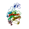
| ||||||||||||
|---|---|---|---|---|---|---|---|---|---|---|---|---|---|
| 1 |
| ||||||||||||
| Unit cell |
| ||||||||||||
| Components on special symmetry positions |
|
-Components
-Protein , 1 types, 1 molecules A
| #1: Protein | Mass: 31614.748 Da / Num. of mol.: 1 / Source method: isolated from a natural source / Source: (natural) Simarouba glauca (aceituno) |
|---|
-Sugars , 2 types, 2 molecules
| #2: Polysaccharide | 2-acetamido-2-deoxy-beta-D-glucopyranose-(1-4)-2-acetamido-2-deoxy-beta-D-glucopyranose-(1-4)-[2- ...2-acetamido-2-deoxy-beta-D-glucopyranose-(1-4)-2-acetamido-2-deoxy-beta-D-glucopyranose-(1-4)-[2-acetamido-2-deoxy-beta-D-glucopyranose-(3-3)]2-acetamido-2-deoxy-beta-D-glucopyranose Type: oligosaccharide / Mass: 830.786 Da / Num. of mol.: 1Source method: isolated from a genetically manipulated source |
|---|---|
| #3: Polysaccharide | 2-acetamido-2-deoxy-beta-D-glucopyranose-(1-4)-2-acetamido-2-deoxy-beta-D-glucopyranose / Mass: 424.401 Da / Num. of mol.: 1 Source method: isolated from a genetically manipulated source |
-Non-polymers , 3 types, 406 molecules 


| #4: Chemical | Chloride Mass: 35.453 Da / Num. of mol.: 3 / Source method: obtained synthetically / Formula: Cl Mass: 35.453 Da / Num. of mol.: 3 / Source method: obtained synthetically / Formula: Cl#5: Chemical |  Mass: 24.305 Da / Num. of mol.: 2 / Source method: obtained synthetically / Formula: Mg Mass: 24.305 Da / Num. of mol.: 2 / Source method: obtained synthetically / Formula: Mg#6: Water | ChemComp-HOH / | WaterMass: 18.015 Da / Num. of mol.: 401 / Source method: isolated from a natural source / Formula: H2O |
|---|
-Details
| Has ligand of interest | Y |
|---|
-Experimental details
-Experiment
| Experiment | Method: X-RAY DIFFRACTION / Number of used crystals: 1 |
|---|
- Sample preparation
| Crystal | Density Matthews: 3.28 Å3/Da / Density % sol: 62.54 % |
|---|---|
| Crystal grow | Temperature: 293 K / Method: vapor diffusion, hanging drop / pH: 8.5 Details: 100mM Tris (pH-8.5), 200mM Magnesium Chloride and 20%PEG |
-Data collection
| Diffraction | Mean temperature: 100 K / Serial crystal experiment: N |
|---|---|
| Diffraction source | Source: SYNCHROTRON / Site: ELETTRA  / Beamline: 11.2C / Wavelength: 1 Å / Beamline: 11.2C / Wavelength: 1 Å |
| Detector | Type: DECTRIS PILATUS 6M / Detector: PIXEL / Date: Sep 21, 2019 / Details: Cilindrical Mirror with 50 nm Pt-coating |
| Radiation | Monochromator: Double Crystal Si111 with LN2 closed loop cooling Protocol: SINGLE WAVELENGTH / Monochromatic (M) / Laue (L): M / Scattering type: x-ray |
| Radiation wavelength | Wavelength: 1 Å / Relative weight: 1 |
| Reflection | Resolution: 1.659→41.482 Å / Num. obs: 49928 / % possible obs: 99.9 % / Redundancy: 6.69 % / Biso Wilson estimate: 18.87 Å2 / CC1/2: 0.999 / Rrim(I) all: 0.076 / Net I/σ(I): 25.5 |
| Reflection shell | Resolution: 1.66→1.7 Å / Mean I/σ(I) obs: 2.35 / Num. unique obs: 7006 / CC1/2: 0.776 / Rrim(I) all: 0.779 / % possible all: 98.2 |
- Processing
| Software |
| |||||||||||||||||||||||||||||||||||||||||||||||||||||||||||||||||||||||||||||||||||||||||||||||||||||||||
|---|---|---|---|---|---|---|---|---|---|---|---|---|---|---|---|---|---|---|---|---|---|---|---|---|---|---|---|---|---|---|---|---|---|---|---|---|---|---|---|---|---|---|---|---|---|---|---|---|---|---|---|---|---|---|---|---|---|---|---|---|---|---|---|---|---|---|---|---|---|---|---|---|---|---|---|---|---|---|---|---|---|---|---|---|---|---|---|---|---|---|---|---|---|---|---|---|---|---|---|---|---|---|---|---|---|---|
| Refinement | Method to determine structure: MOLECULAR REPLACEMENT Starting model: 4MST Resolution: 1.66→36.72 Å / SU ML: 0.1772 / Cross valid method: FREE R-VALUE / σ(F): 1.35 / Phase error: 17.8709 Stereochemistry target values: GeoStd + Monomer Library + CDL v1.2
| |||||||||||||||||||||||||||||||||||||||||||||||||||||||||||||||||||||||||||||||||||||||||||||||||||||||||
| Solvent computation | Shrinkage radii: 0.9 Å / VDW probe radii: 1.11 Å / Solvent model: FLAT BULK SOLVENT MODEL | |||||||||||||||||||||||||||||||||||||||||||||||||||||||||||||||||||||||||||||||||||||||||||||||||||||||||
| Displacement parameters | Biso mean: 24.39 Å2 | |||||||||||||||||||||||||||||||||||||||||||||||||||||||||||||||||||||||||||||||||||||||||||||||||||||||||
| Refinement step | Cycle: LAST / Resolution: 1.66→36.72 Å
| |||||||||||||||||||||||||||||||||||||||||||||||||||||||||||||||||||||||||||||||||||||||||||||||||||||||||
| Refine LS restraints |
| |||||||||||||||||||||||||||||||||||||||||||||||||||||||||||||||||||||||||||||||||||||||||||||||||||||||||
| LS refinement shell |
|
