 Movie
Movie Controller

[English] 日本語
 Yorodumi
- PDB-6dnd: Crystal structure of wild-type (WT) human Glutamate oxaloacetate ... -
+ Open data

- Basic information
| Entry | Database: PDB / ID: 6dnd | ||||||
|---|---|---|---|---|---|---|---|
| Title | Crystal structure of wild-type (WT) human Glutamate oxaloacetate transaminase 1 (GOT1) | ||||||
 Components Components | Aspartate aminotransferase, cytoplasmic Aspartate transaminase Aspartate transaminase | ||||||
 Keywords Keywords | TRANSFERASE / Aspartate aminotransferase / Glutamate oxaloacetate transaminase 1 / GOT1 / PLP | ||||||
| Function / homology |  Function and homology informationphosphatidylserine decarboxylase activity / glutamate catabolic process to aspartate / glutamate catabolic process to 2-oxoglutarate / cysteine transaminase / L-cysteine transaminase activity / Methionine salvage pathway / aspartate biosynthetic process / glycerol biosynthetic process / aspartate metabolic process / Aspartate and asparagine metabolism ...phosphatidylserine decarboxylase activity / glutamate catabolic process to aspartate / glutamate catabolic process to 2-oxoglutarate / cysteine transaminase / L-cysteine transaminase activity / Methionine salvage pathway / aspartate biosynthetic process / glycerol biosynthetic process / aspartate metabolic process / Aspartate and asparagine metabolism / glutamate metabolic process / aspartate catabolic process / 2-oxoglutarate metabolic process / aspartate transaminase / oxaloacetate metabolic process / L-aspartate:2-oxoglutarate aminotransferase activity / Gluconeogenesis / fatty acid homeostasis / response to glucocorticoid / Notch signaling pathway / gluconeogenesis / cellular response to insulin stimulus / pyridoxal phosphate binding / extracellular exosome / nucleus / cytosol / cytoplasm Function and homology informationphosphatidylserine decarboxylase activity / glutamate catabolic process to aspartate / glutamate catabolic process to 2-oxoglutarate / cysteine transaminase / L-cysteine transaminase activity / Methionine salvage pathway / aspartate biosynthetic process / glycerol biosynthetic process / aspartate metabolic process / Aspartate and asparagine metabolism ...phosphatidylserine decarboxylase activity / glutamate catabolic process to aspartate / glutamate catabolic process to 2-oxoglutarate / cysteine transaminase / L-cysteine transaminase activity / Methionine salvage pathway / aspartate biosynthetic process / glycerol biosynthetic process / aspartate metabolic process / Aspartate and asparagine metabolism / glutamate metabolic process / aspartate catabolic process / 2-oxoglutarate metabolic process / aspartate transaminase / oxaloacetate metabolic process / L-aspartate:2-oxoglutarate aminotransferase activity / Gluconeogenesis / fatty acid homeostasis / response to glucocorticoid / Notch signaling pathway / gluconeogenesis / cellular response to insulin stimulus / pyridoxal phosphate binding / extracellular exosome / nucleus / cytosol / cytoplasmSimilarity search - Function | ||||||
| Biological species |  Homo sapiens (human) Homo sapiens (human) | ||||||
| Method | X-RAY DIFFRACTION / SYNCHROTRON / MOLECULAR REPLACEMENT / Resolution: 2.1 Å | ||||||
 Authors Authors | Assar, Z. / Holt, M.C. / Stein, A.J. / Lairson, L. / Lyssiotis, C.A. | ||||||
 Citation Citation | Journal: Biochemistry / Year: 2018 Title: Biochemical Characterization and Structure-Based Mutational Analysis Provide Insight into the Binding and Mechanism of Action of Novel Aspartate Aminotransferase Inhibitors. Authors: Holt, M.C. / Assar, Z. / Beheshti Zavareh, R. / Lin, L. / Anglin, J. / Mashadova, O. / Haldar, D. / Mullarky, E. / Kremer, D.M. / Cantley, L.C. / Kimmelman, A.C. / Stein, A.J. / Lairson, L.L. / Lyssiotis, C.A. | ||||||
| History |
|
- Structure visualization
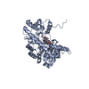
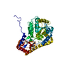
| Structure viewer | Molecule: MolmilJmol/JSmol |
|---|
- Downloads & links
-Download
| PDBx/mmCIF format | 6dnd.cif.gz | 175.1 KB | Display | PDBx/mmCIF format |
|---|---|---|---|---|
| PDB format | pdb6dnd.ent.gz | 138.4 KB | Display | PDB format |
| PDBx/mmJSON format | 6dnd.json.gz | Tree view | PDBx/mmJSON format | |
| Others |  Other downloads Other downloads |
-Validation report
| Arichive directory | https://data.pdbj.org/pub/pdb/validation_reports/dn/6dndftp://data.pdbj.org/pub/pdb/validation_reports/dn/6dnd | HTTPS FTP |
|---|
-Related structure data
| Related structure data |  6dnaC  6dnbC  3ii0S S: Starting model for refinement C: citing same article ( |
|---|---|
| Similar structure data |


-Links
 PDBj
PDBj
- Assembly
| Deposited unit | 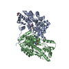
| ||||||||||||||||||
|---|---|---|---|---|---|---|---|---|---|---|---|---|---|---|---|---|---|---|---|
| 1 | 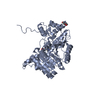
| ||||||||||||||||||
| 2 | 
| ||||||||||||||||||
| Unit cell |
| ||||||||||||||||||
| Noncrystallographic symmetry (NCS) | NCS domain:
NCS domain segments: Component-ID: 0 / Ens-ID: 1 / Beg auth comp-ID: PRO / Beg label comp-ID: PRO / End auth comp-ID: THR / End label comp-ID: THR / Refine code: 0 / Auth seq-ID: 3 - 410 / Label seq-ID: 1 - 408
|
-Components
| #1: Protein | Aspartate transaminase / cAspAT / Cysteine aminotransferase / cytoplasmic / Cysteine transaminase / cCAT / Glutamate ...cAspAT / Cysteine aminotransferase / cytoplasmic / Cysteine transaminase / cCAT / Glutamate oxaloacetate transaminase 1 / Transaminase A Mass: 46097.039 Da / Num. of mol.: 2 Source method: isolated from a genetically manipulated source Source: (gene. exp.) Homo sapiens (human) / Gene: GOT1 / Production host:  Escherichia coli (E. coli) Escherichia coli (E. coli)References: UniProt: P17174, aspartate transaminase, cysteine transaminase#2: Chemical | Polyethylene glycol  Mass: 194.226 Da / Num. of mol.: 2 / Source method: obtained synthetically / Formula: C8H18O5 / Comment: precipitant*YM Mass: 194.226 Da / Num. of mol.: 2 / Source method: obtained synthetically / Formula: C8H18O5 / Comment: precipitant*YM#3: Chemical | Pyridoxal phosphate  Mass: 247.142 Da / Num. of mol.: 2 / Source method: obtained synthetically / Formula: C8H10NO6P Mass: 247.142 Da / Num. of mol.: 2 / Source method: obtained synthetically / Formula: C8H10NO6P#4: Water | ChemComp-HOH / | Water Mass: 18.015 Da / Num. of mol.: 101 / Source method: isolated from a natural source / Formula: H2O Mass: 18.015 Da / Num. of mol.: 101 / Source method: isolated from a natural source / Formula: H2O |
|---|
-Experimental details
-Experiment
| Experiment | Method: X-RAY DIFFRACTION / Number of used crystals: 1 |
|---|
- Sample preparation
| Crystal | Density Matthews: 2.35 Å3/Da / Density % sol: 47.64 % |
|---|---|
| Crystal grow | Temperature: 298 K / Method: vapor diffusion, sitting drop Details: 0.1 M Sodium Acetate Trihydrate pH 4.5, 25% w/v PEG 3,350 |
-Data collection
| Diffraction | Mean temperature: 100 K |
|---|---|
| Diffraction source | Source: SYNCHROTRON / Site: APS  / Beamline: 21-ID-D / Wavelength: 0.9787 Å / Beamline: 21-ID-D / Wavelength: 0.9787 Å |
| Detector | Type: DECTRIS EIGER X 9M / Detector: PIXEL / Date: Jul 21, 2016 |
| Radiation | Protocol: SINGLE WAVELENGTH / Monochromatic (M) / Laue (L): M / Scattering type: x-ray |
| Radiation wavelength | Wavelength: 0.9787 Å / Relative weight: 1 |
| Reflection | Resolution: 2.1→50 Å / Num. obs: 3512 / % possible obs: 99.92 % / Redundancy: 8 % / Rmerge(I) obs: 0.07 / Net I/σ(I): 30.01 |
| Reflection shell | Resolution: 2.1→2.15 Å / Rmerge(I) obs: 0.32 |
- Processing
| Software |
| ||||||||||||||||||||||||||||||||||||||||||||||||||||||||||||||||||||||||||||||||||||||||||||||||||||||||||||||||||||||||||||||||||||||||||||||||||||||||||||||||||||||||||||||||||||||
|---|---|---|---|---|---|---|---|---|---|---|---|---|---|---|---|---|---|---|---|---|---|---|---|---|---|---|---|---|---|---|---|---|---|---|---|---|---|---|---|---|---|---|---|---|---|---|---|---|---|---|---|---|---|---|---|---|---|---|---|---|---|---|---|---|---|---|---|---|---|---|---|---|---|---|---|---|---|---|---|---|---|---|---|---|---|---|---|---|---|---|---|---|---|---|---|---|---|---|---|---|---|---|---|---|---|---|---|---|---|---|---|---|---|---|---|---|---|---|---|---|---|---|---|---|---|---|---|---|---|---|---|---|---|---|---|---|---|---|---|---|---|---|---|---|---|---|---|---|---|---|---|---|---|---|---|---|---|---|---|---|---|---|---|---|---|---|---|---|---|---|---|---|---|---|---|---|---|---|---|---|---|---|---|
| Refinement | Method to determine structure: MOLECULAR REPLACEMENT Starting model: 3ii0 Resolution: 2.1→68.85 Å / Cor.coef. Fo:Fc: 0.962 / Cor.coef. Fo:Fc free: 0.931 / SU B: 7.27 / SU ML: 0.186 / Cross valid method: THROUGHOUT / ESU R: 0.243 / ESU R Free: 0.212 / Stereochemistry target values: MAXIMUM LIKELIHOOD / Details: HYDROGENS HAVE BEEN ADDED IN THE RIDING POSITIONS
| ||||||||||||||||||||||||||||||||||||||||||||||||||||||||||||||||||||||||||||||||||||||||||||||||||||||||||||||||||||||||||||||||||||||||||||||||||||||||||||||||||||||||||||||||||||||
| Solvent computation | Ion probe radii: 0.8 Å / Shrinkage radii: 0.8 Å / VDW probe radii: 1.2 Å / Solvent model: MASK | ||||||||||||||||||||||||||||||||||||||||||||||||||||||||||||||||||||||||||||||||||||||||||||||||||||||||||||||||||||||||||||||||||||||||||||||||||||||||||||||||||||||||||||||||||||||
| Displacement parameters | Biso mean: 57.577 Å2
| ||||||||||||||||||||||||||||||||||||||||||||||||||||||||||||||||||||||||||||||||||||||||||||||||||||||||||||||||||||||||||||||||||||||||||||||||||||||||||||||||||||||||||||||||||||||
| Refinement step | Cycle: 1 / Resolution: 2.1→68.85 Å
| ||||||||||||||||||||||||||||||||||||||||||||||||||||||||||||||||||||||||||||||||||||||||||||||||||||||||||||||||||||||||||||||||||||||||||||||||||||||||||||||||||||||||||||||||||||||
| Refine LS restraints |
|