 Movie
Movie Controller

[English] 日本語
 Yorodumi
- PDB-1akb: STRUCTURAL BASIS FOR THE CATALYTIC ACTIVITY OF ASPARTATE AMINOTRA... -
+ Open data

- Basic information
| Entry | Database: PDB / ID: 1akb | ||||||
|---|---|---|---|---|---|---|---|
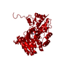
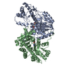
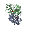
| Title | STRUCTURAL BASIS FOR THE CATALYTIC ACTIVITY OF ASPARTATE AMINOTRANSFERASE K258H LACKING ITS PYRIDOXAL-5'-PHOSPHATE-BINDING LYSINE RESIDUE | ||||||
 Components Components | ASPARTATE AMINOTRANSFERASE Aspartate transaminase Aspartate transaminase | ||||||
 Keywords Keywords | TRANSFERASE(AMINOTRANSFERASE) | ||||||
| Function / homology |  Function and homology informationAmino acid metabolism / Gluconeogenesis / kynurenine-oxoglutarate transaminase / kynurenine-oxoglutarate transaminase activity / aspartate metabolic process / glutamate metabolic process / L-phenylalanine biosynthetic process from chorismate via phenylpyruvate / L-tyrosine:2-oxoglutarate aminotransferase activity / aspartate catabolic process / 2-oxoglutarate metabolic process ...Amino acid metabolism / Gluconeogenesis / kynurenine-oxoglutarate transaminase / kynurenine-oxoglutarate transaminase activity / aspartate metabolic process / glutamate metabolic process / L-phenylalanine biosynthetic process from chorismate via phenylpyruvate / L-tyrosine:2-oxoglutarate aminotransferase activity / aspartate catabolic process / 2-oxoglutarate metabolic process / aspartate transaminase / L-aspartate:2-oxoglutarate aminotransferase activity / pyridoxal phosphate binding / mitochondrial matrix / protein homodimerization activity / mitochondrion / cytosol Function and homology informationAmino acid metabolism / Gluconeogenesis / kynurenine-oxoglutarate transaminase / kynurenine-oxoglutarate transaminase activity / aspartate metabolic process / glutamate metabolic process / L-phenylalanine biosynthetic process from chorismate via phenylpyruvate / L-tyrosine:2-oxoglutarate aminotransferase activity / aspartate catabolic process / 2-oxoglutarate metabolic process ...Amino acid metabolism / Gluconeogenesis / kynurenine-oxoglutarate transaminase / kynurenine-oxoglutarate transaminase activity / aspartate metabolic process / glutamate metabolic process / L-phenylalanine biosynthetic process from chorismate via phenylpyruvate / L-tyrosine:2-oxoglutarate aminotransferase activity / aspartate catabolic process / 2-oxoglutarate metabolic process / aspartate transaminase / L-aspartate:2-oxoglutarate aminotransferase activity / pyridoxal phosphate binding / mitochondrial matrix / protein homodimerization activity / mitochondrion / cytosolSimilarity search - Function | ||||||
| Biological species |  Gallus gallus (chicken) Gallus gallus (chicken) | ||||||
| Method | X-RAY DIFFRACTION / Resolution: 2.3 Å | ||||||
 Authors Authors | Malashkevich, V.N. / Jansonius, J.N. | ||||||
 Citation Citation | Journal: Biochemistry / Year: 1995 Title: Structural basis for the catalytic activity of aspartate aminotransferase K258H lacking the pyridoxal 5'-phosphate-binding lysine residue. Authors: Malashkevich, V.N. / Jager, J. / Ziak, M. / Sauder, U. / Gehring, H. / Christen, P. / Jansonius, J.N. #1: Journal: Eur.J.Biochem. / Year: 1993Title: Mutant Aspartate Aminotransferase (K258H) without Pyridoxal-5'-Phosphate-Binding Lysine Residue. Structural and Catalytic Properties Authors: Ziak, M. / Jaeger, J. / Malashkevich, V.N. / Jaussi, R. / Gehring, H. / Jansonius, J.N. / Christen, P. #2: Journal: Biological Macromolecules and Assemblies / Year: 1987Title: Structural Basis for Catalysis by Aspartate Aminotransferase Authors: Jansonius, J.N. / Vincent, M.G. | ||||||
| History |
|
- Structure visualization
| Structure viewer | Molecule: MolmilJmol/JSmol |
|---|
- Downloads & links
-Download
| PDBx/mmCIF format | 1akb.cif.gz | 100.6 KB | Display | PDBx/mmCIF format |
|---|---|---|---|---|
| PDB format | pdb1akb.ent.gz | 76.2 KB | Display | PDB format |
| PDBx/mmJSON format | 1akb.json.gz | Tree view | PDBx/mmJSON format | |
| Others |  Other downloads Other downloads |
-Validation report
| Arichive directory | https://data.pdbj.org/pub/pdb/validation_reports/ak/1akbftp://data.pdbj.org/pub/pdb/validation_reports/ak/1akb | HTTPS FTP |
|---|
-Related structure data


-Links
 PDBj
PDBj- Assembly
| Deposited unit | 
| ||||||||
|---|---|---|---|---|---|---|---|---|---|
| 1 | 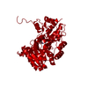
| ||||||||
| Unit cell |
| ||||||||
| Atom site foot note | 1: CIS PROLINE - PRO A 138 / 2: CIS PROLINE - PRO A 195 3: K258H ACTIVE SITE MUTANT. THE RESIDUE PPD A 411 REPRESENTS THE COVALENT ADDUCT BETWEEN SUBSTRATE L-ASPARTATE AND PYRIDOXAL-5'-PHOSPHATE WITH THE REDUCED ALDIMINE BOND. |
-Components
| #1: Protein | Aspartate transaminase Mass: 45001.453 Da / Num. of mol.: 1 / Mutation: K250H Source method: isolated from a genetically manipulated source Source: (gene. exp.) Gallus gallus (chicken) / Organ: HEART / Production host: unidentified (others) / References: UniProt: P00508, aspartate transaminase | ||
|---|---|---|---|
| #2: Chemical | ChemComp-PPD /   Mass: 364.245 Da / Num. of mol.: 1 / Source method: obtained synthetically / Formula: C12H17N2O9P Mass: 364.245 Da / Num. of mol.: 1 / Source method: obtained synthetically / Formula: C12H17N2O9P | ||
| #3: Water | ChemComp-HOH / Water Mass: 18.015 Da / Num. of mol.: 312 / Source method: isolated from a natural source / Formula: H2O Mass: 18.015 Da / Num. of mol.: 312 / Source method: isolated from a natural source / Formula: H2O | ||
| Nonpolymer details | ONE MOLECULE OF N-(5'-PHOSPHOPYR| Sequence details | THE RESIDUES ARE NUMBERED FROM 3 - 410, ACCORDING TO A SEQUENCE ALIGNMENT WITH THE LONGER SEQUENCE ...THE RESIDUES ARE NUMBERED FROM 3 - 410, ACCORDING TO A SEQUENCE ALIGNMENT WITH THE LONGER SEQUENCE OF PIG CYTOSOLIC ASPARTATE AMINOTRANS | |
-Experimental details
-Experiment
| Experiment | Method: X-RAY DIFFRACTION |
|---|
- Sample preparation
| Crystal | Density Matthews: 2.3 Å3/Da / Density % sol: 46.64 % | ||||||||||||||||||||||||||||||||||||||||||||||||
|---|---|---|---|---|---|---|---|---|---|---|---|---|---|---|---|---|---|---|---|---|---|---|---|---|---|---|---|---|---|---|---|---|---|---|---|---|---|---|---|---|---|---|---|---|---|---|---|---|---|
| Crystal grow | *PLUS pH: 7.5 / Method: other | ||||||||||||||||||||||||||||||||||||||||||||||||
| Components of the solutions | *PLUS
|
-Data collection
| Radiation | Scattering type: x-ray |
|---|---|
| Radiation wavelength | Relative weight: 1 |
| Reflection | *PLUS Highest resolution: 2.3 Å / Lowest resolution: 10 Å / Num. obs: 18825 / % possible obs: 96.2 % / Num. measured all: 72346 / Rmerge(I) obs: 0.082 |
- Processing
| Software | Name: PROLSQ / Classification: refinement | ||||||||||||||||||||||||||||||||||||||||||||||||||||||||||||||||||||||||||||||||||||
|---|---|---|---|---|---|---|---|---|---|---|---|---|---|---|---|---|---|---|---|---|---|---|---|---|---|---|---|---|---|---|---|---|---|---|---|---|---|---|---|---|---|---|---|---|---|---|---|---|---|---|---|---|---|---|---|---|---|---|---|---|---|---|---|---|---|---|---|---|---|---|---|---|---|---|---|---|---|---|---|---|---|---|---|---|---|
| Refinement | Resolution: 2.3→10 Å / σ(F): 1 /
| ||||||||||||||||||||||||||||||||||||||||||||||||||||||||||||||||||||||||||||||||||||
| Refinement step | Cycle: LAST / Resolution: 2.3→10 Å
| ||||||||||||||||||||||||||||||||||||||||||||||||||||||||||||||||||||||||||||||||||||
| Refine LS restraints |
| ||||||||||||||||||||||||||||||||||||||||||||||||||||||||||||||||||||||||||||||||||||
| Software | *PLUS Name: PROLSQ / Classification: refinement | ||||||||||||||||||||||||||||||||||||||||||||||||||||||||||||||||||||||||||||||||||||
| Refinement | *PLUS Rfactor obs: 0.178 | ||||||||||||||||||||||||||||||||||||||||||||||||||||||||||||||||||||||||||||||||||||
| Solvent computation | *PLUS | ||||||||||||||||||||||||||||||||||||||||||||||||||||||||||||||||||||||||||||||||||||
| Displacement parameters | *PLUS |
