 Movie
Movie Controller

[English] 日本語
 Yorodumi
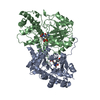
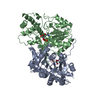
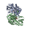
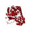
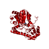
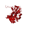
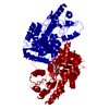
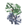
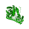
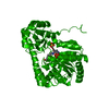
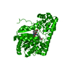
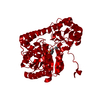
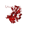
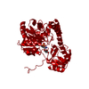
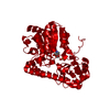
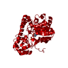
- PDB-1czc: ASPARTATE AMINOTRANSFERASE MUTANT ATB17/139S/142N WITH GLUTARIC ACID -
+ Open data

- Basic information
| Entry | Database: PDB / ID: 1czc | ||||||
|---|---|---|---|---|---|---|---|
| Title | ASPARTATE AMINOTRANSFERASE MUTANT ATB17/139S/142N WITH GLUTARIC ACID | ||||||
 Components Components | PROTEIN (ASPARTATE AMINOTRANSFERASE) | ||||||
 Keywords Keywords |  TRANSFERASE / ASPARTATE AMINOTRANSFERASE / SUBSTRATE SPECIFICITY TRANSFERASE / ASPARTATE AMINOTRANSFERASE / SUBSTRATE SPECIFICITY | ||||||
| Function / homology |  Function and homology information Function and homology informationL-phenylalanine biosynthetic process from chorismate via phenylpyruvate / L-phenylalanine biosynthetic process / L-tyrosine:2-oxoglutarate aminotransferase activity / aspartate catabolic process / aspartate transaminase / L-aspartate:2-oxoglutarate aminotransferase activity / pyridoxal phosphate binding / protein homodimerization activity / identical protein binding / cytosol / cytoplasmSimilarity search - Function | ||||||
| Biological species |  Escherichia coli (E. coli) Escherichia coli (E. coli) | ||||||
| Method | X-RAY DIFFRACTION / MOLECULAR REPLACEMENT / Resolution: 2.5 Å | ||||||
 Authors Authors | Okamoto, A. / Oue, S. / Yano, T. / Kagamiyama, H. | ||||||
 Citation Citation | Journal: J.Biochem.(Tokyo) / Year: 2000 Title: Cocrystallization of a mutant aspartate aminotransferase with a C5-dicarboxylic substrate analog: structural comparison with the enzyme-C4-dicarboxylic analog complex. Authors: Oue, S. / Okamoto, A. / Yano, T. / Kagamiyama, H. #1: Journal: J.Biol.Chem. / Year: 1999Title: Redesigning the Substrate Specificity of an Enzyme by Cumulative Effects of the Mutations of Non-Active Site Residues Authors: Oue, S. / Okamoto, A. / Yano, T. / Kagamiyama, H. #2: Journal: Proc.Natl.Acad.Sci.USA / Year: 1998Title: Directed Evolution of an Aspartate Aminotransferase with New Substrate Specificities Authors: Yano, T. / Oue, S. / Kagamiyama, H. | ||||||
| History |
|
- Structure visualization
| Structure viewer | Molecule: MolmilJmol/JSmol |
|---|
- Downloads & links
-Download
| PDBx/mmCIF format | 1czc.cif.gz | 94.2 KB | Display | PDBx/mmCIF format |
|---|---|---|---|---|
| PDB format | pdb1czc.ent.gz | 69.7 KB | Display | PDB format |
| PDBx/mmJSON format | 1czc.json.gz | Tree view | PDBx/mmJSON format | |
| Others |  Other downloads Other downloads |
-Validation report
| Arichive directory | https://data.pdbj.org/pub/pdb/validation_reports/cz/1czcftp://data.pdbj.org/pub/pdb/validation_reports/cz/1czc | HTTPS FTP |
|---|
-Related structure data
| Related structure data |  1czeC  1yooS S: Starting model for refinement C: citing same article ( |
|---|---|
| Similar structure data |

-Links
 PDBj
PDBj- Assembly
| Deposited unit | 
| ||||||||||
|---|---|---|---|---|---|---|---|---|---|---|---|
| 1 | 
| ||||||||||
| Unit cell |
|
-Components
| #1: Protein | Mass: 43662.207 Da / Num. of mol.: 1 Mutation: A11T,F24L,N34D,I37M,K41N,K126R,A269T,A293V, N297S,S311G,I353T,S361F,S363G, V387L,M397L Source method: isolated from a genetically manipulated source Source: (gene. exp.) Escherichia coli (E. coli) / Plasmid: PUC118 / Production host: Escherichia coli (E. coli) / Strain (production host): TY103 / References: UniProt: P00509, aspartate transaminase |
|---|---|
| #2: Chemical | ChemComp-PLP / Pyridoxal phosphate  Mass: 247.142 Da / Num. of mol.: 1 / Source method: obtained synthetically / Formula: C8H10NO6P Mass: 247.142 Da / Num. of mol.: 1 / Source method: obtained synthetically / Formula: C8H10NO6P |
| #3: Chemical | ChemComp-GUA / Glutaric acid  Mass: 132.115 Da / Num. of mol.: 1 / Source method: obtained synthetically / Formula: C5H8O4 Mass: 132.115 Da / Num. of mol.: 1 / Source method: obtained synthetically / Formula: C5H8O4 |
| #4: Water | ChemComp-HOH / Water Mass: 18.015 Da / Num. of mol.: 160 / Source method: isolated from a natural source / Formula: H2O Mass: 18.015 Da / Num. of mol.: 160 / Source method: isolated from a natural source / Formula: H2O |
-Experimental details
-Experiment
| Experiment | Method: X-RAY DIFFRACTION / Number of used crystals: 1 |
|---|
- Sample preparation
| Crystal | Density Matthews: 2.99 Å3/Da / Density % sol: 58.9 % | ||||||||||||||||||||||||||||||
|---|---|---|---|---|---|---|---|---|---|---|---|---|---|---|---|---|---|---|---|---|---|---|---|---|---|---|---|---|---|---|---|
| Crystal grow | Temperature: 293 K / Method: vapor diffusion, sitting drop / pH: 7.5 Details: PROTEIN WAS CRYSTALLIZED FROM 1.7M AMMONIUM SULFATE, 0.1 M SODIUM HEPES, 0.167 M GLUTARIC ACID, pH 7.5, VAPOR DIFFUSION, SITTING DROP, temperature 293.0K | ||||||||||||||||||||||||||||||
| Crystal grow | *PLUS Temperature: 20 ℃ | ||||||||||||||||||||||||||||||
| Components of the solutions | *PLUS
|
-Data collection
| Diffraction | Mean temperature: 293 K |
|---|---|
| Diffraction source | Source: ROTATING ANODE / Type: RIGAKU RU200 / Wavelength: 1.5418 |
| Detector | Type: RIGAKU RAXIS IIC / Detector: IMAGE PLATE / Date: Feb 15, 1999 / Details: MIRROR |
| Radiation | Protocol: SINGLE WAVELENGTH / Monochromatic (M) / Laue (L): M / Scattering type: x-ray |
| Radiation wavelength | Wavelength: 1.5418 Å / Relative weight: 1 |
| Reflection | Resolution: 2.5→75 Å / Num. obs: 16658 / % possible obs: 98.8 % / Observed criterion σ(I): 1 / Redundancy: 3.7 % / Biso Wilson estimate: 23.5 Å2 / Rmerge(I) obs: 0.0536 / Net I/σ(I): 16.7 |
| Reflection shell | Resolution: 2.5→2.8 Å / Redundancy: 2.7 % / Rmerge(I) obs: 0.088 / Mean I/σ(I) obs: 64 / % possible all: 97.5 |
| Reflection | *PLUS Num. measured all: 61781 |
| Reflection shell | *PLUS % possible obs: 97.5 % |
- Processing
| Software |
| ||||||||||||||||||||||||||||||||||||||||||||||||||||||||||||||||||||||||||||||||
|---|---|---|---|---|---|---|---|---|---|---|---|---|---|---|---|---|---|---|---|---|---|---|---|---|---|---|---|---|---|---|---|---|---|---|---|---|---|---|---|---|---|---|---|---|---|---|---|---|---|---|---|---|---|---|---|---|---|---|---|---|---|---|---|---|---|---|---|---|---|---|---|---|---|---|---|---|---|---|---|---|---|
| Refinement | Method to determine structure: MOLECULAR REPLACEMENT Starting model: 1YOO Resolution: 2.5→10 Å / Rfactor Rfree error: 0.006 / Data cutoff high absF: 10000000 / Data cutoff low absF: 0 / Isotropic thermal model: RESTRAINED / Cross valid method: THROUGHOUT / σ(F): 2 / Stereochemistry target values: ENGH & HUBER
| ||||||||||||||||||||||||||||||||||||||||||||||||||||||||||||||||||||||||||||||||
| Displacement parameters | Biso mean: 21.1 Å2 | ||||||||||||||||||||||||||||||||||||||||||||||||||||||||||||||||||||||||||||||||
| Refine analyze |
| ||||||||||||||||||||||||||||||||||||||||||||||||||||||||||||||||||||||||||||||||
| Refinement step | Cycle: LAST / Resolution: 2.5→10 Å
| ||||||||||||||||||||||||||||||||||||||||||||||||||||||||||||||||||||||||||||||||
| Refine LS restraints |
| ||||||||||||||||||||||||||||||||||||||||||||||||||||||||||||||||||||||||||||||||
| LS refinement shell | Resolution: 2.5→2.65 Å / Rfactor Rfree error: 0.018 / Total num. of bins used: 6
| ||||||||||||||||||||||||||||||||||||||||||||||||||||||||||||||||||||||||||||||||
| Xplor file |
| ||||||||||||||||||||||||||||||||||||||||||||||||||||||||||||||||||||||||||||||||
| Software | *PLUS Name: X-PLOR / Version: 3.851 / Classification: refinement | ||||||||||||||||||||||||||||||||||||||||||||||||||||||||||||||||||||||||||||||||
| Refine LS restraints | *PLUS
|
