 Movie
Movie Controller


+ Open data

- Basic information
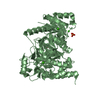
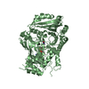
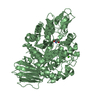
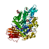
| Entry | Database: PDB / ID: 6dk2 | ||||||
|---|---|---|---|---|---|---|---|
| Title | Bacteroidetes AC2a SusD-like | ||||||
 Components Components | SusD Lunca, Bihor Lunca, Bihor | ||||||
 Keywords Keywords | SUGAR BINDING PROTEIN / SusD-like / cellulose / rumen / Bacteroidetes | ||||||
| Function / homology | SusD-like 2 / Starch-binding associating with outer membrane / Tetratricopeptide-like helical domain superfamily / SusD Function and homology information Function and homology information | ||||||
| Biological species |  Bacteroidetes bacterium AC2a (bacteria) Bacteroidetes bacterium AC2a (bacteria) | ||||||
| Method | X-RAY DIFFRACTION / SYNCHROTRON / MOLECULAR REPLACEMENT / molecular replacement / Resolution: 2.02 Å | ||||||
 Authors Authors | Koropatkin, N.M. | ||||||
 Citation Citation | Journal: To Be Published Title: To Be Published Authors: Koropatkin, N.M. / Bahr, C.M.E. / Pope, P.B. / Naas, A.E. | ||||||
| History |
|
- Structure visualization
| Structure viewer | Molecule: MolmilJmol/JSmol |
|---|
- Downloads & links
-Download
| PDBx/mmCIF format | 6dk2.cif.gz | 477.5 KB | Display | PDBx/mmCIF format |
|---|---|---|---|---|
| PDB format | pdb6dk2.ent.gz | 391.1 KB | Display | PDB format |
| PDBx/mmJSON format | 6dk2.json.gz | Tree view | PDBx/mmJSON format | |
| Others |  Other downloads Other downloads |
-Validation report
| Arichive directory | https://data.pdbj.org/pub/pdb/validation_reports/dk/6dk2ftp://data.pdbj.org/pub/pdb/validation_reports/dk/6dk2 | HTTPS FTP |
|---|
-Related structure data
| Related structure data | 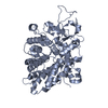 5j5uS S: Starting model for refinement |
|---|---|
| Similar structure data |


-Links
 PDBj
PDBj- Assembly
| Deposited unit | 
| ||||||||
|---|---|---|---|---|---|---|---|---|---|
| 1 | 
| ||||||||
| 2 | 
| ||||||||
| Unit cell |
|
-Components
| #1: Protein | Lunca, Bihor Mass: 59657.348 Da / Num. of mol.: 2 / Fragment: UNP residues 33-560 Source method: isolated from a genetically manipulated source Details: metagenomic data set Source: (gene. exp.) Bacteroidetes bacterium AC2a (bacteria)Plasmid: PETite Nhis / Production host: Escherichia coli (E. coli) / Strain (production host): Rosetta(DE3)pLysS / References: UniProt: A0A076ML24#2: Chemical | ChemComp-SO4 / Sulfate  Mass: 96.063 Da / Num. of mol.: 14 / Source method: obtained synthetically / Formula: SO4 Mass: 96.063 Da / Num. of mol.: 14 / Source method: obtained synthetically / Formula: SO4#3: Chemical | ChemComp-EDO / Ethylene glycol  Mass: 62.068 Da / Num. of mol.: 28 / Source method: obtained synthetically / Formula: C2H6O2 Mass: 62.068 Da / Num. of mol.: 28 / Source method: obtained synthetically / Formula: C2H6O2#4: Chemical | Chloride  Mass: 35.453 Da / Num. of mol.: 2 / Source method: isolated from a natural source / Formula: Cl Mass: 35.453 Da / Num. of mol.: 2 / Source method: isolated from a natural source / Formula: Cl#5: Water | ChemComp-HOH / | Water Mass: 18.015 Da / Num. of mol.: 984 / Source method: isolated from a natural source / Formula: H2O Mass: 18.015 Da / Num. of mol.: 984 / Source method: isolated from a natural source / Formula: H2O |
|---|
-Experimental details
-Experiment
| Experiment | Method: X-RAY DIFFRACTION / Number of used crystals: 1 |
|---|
- Sample preparation
| Crystal grow | Temperature: 277 K / Method: vapor diffusion, hanging drop / pH: 7.5 / Details: 1.5 M ammonium sulphate, 50 mM Tris-HCl pH 7.5 / Temp details: cold room |
|---|
-Data collection
| Diffraction | Mean temperature: 100 K / Ambient temp details: cryo |
|---|---|
| Diffraction source | Source: SYNCHROTRON / Site: APS  / Beamline: 21-ID-F / Wavelength: 0.979 Å / Beamline: 21-ID-F / Wavelength: 0.979 Å |
| Detector | Type: RAYONIX MX-300 / Detector: CCD / Date: Nov 15, 2015 |
| Radiation | Monochromator: C(111) / Protocol: SINGLE WAVELENGTH / Monochromatic (M) / Laue (L): M / Scattering type: x-ray |
| Radiation wavelength | Wavelength: 0.979 Å / Relative weight: 1 |
| Reflection | Resolution: 2.02→56.93 Å / Num. obs: 172008 / % possible obs: 99.71 % / Redundancy: 6.2 % / Biso Wilson estimate: 35.74 Å2 / CC1/2: 0.997 / Rmerge(I) obs: 0.085 / Rrim(I) all: 0.093 / Net I/σ(I): 11.13 |
| Reflection shell | Resolution: 2.02→2.094 Å / Redundancy: 6.2 % / Rmerge(I) obs: 0.521 / Mean I/σ(I) obs: 2.27 / Num. unique obs: 17235 / CC1/2: 0.914 / % possible all: 99.99 |
-Phasing
| Phasing | Method: molecular replacement | |||||||||
|---|---|---|---|---|---|---|---|---|---|---|
| Phasing MR |
|
- Processing
| Software |
| |||||||||||||||||||||||||||||||||||||||||||||||||||||||||||||||||||||||||||
|---|---|---|---|---|---|---|---|---|---|---|---|---|---|---|---|---|---|---|---|---|---|---|---|---|---|---|---|---|---|---|---|---|---|---|---|---|---|---|---|---|---|---|---|---|---|---|---|---|---|---|---|---|---|---|---|---|---|---|---|---|---|---|---|---|---|---|---|---|---|---|---|---|---|---|---|---|
| Refinement | Method to determine structure: MOLECULAR REPLACEMENT Starting model: 5J5U Resolution: 2.02→56.93 Å / Cor.coef. Fo:Fc: 0.972 / Cor.coef. Fo:Fc free: 0.964 / SU B: 5.489 / SU ML: 0.065 / Cross valid method: THROUGHOUT / σ(F): 0 / ESU R: 0.108 / ESU R Free: 0.087 Details: HYDROGENS HAVE BEEN ADDED IN THE RIDING POSITIONS U VALUES : WITH TLS ADDED
| |||||||||||||||||||||||||||||||||||||||||||||||||||||||||||||||||||||||||||
| Solvent computation | Ion probe radii: 0.8 Å / Shrinkage radii: 0.8 Å / VDW probe radii: 1.2 Å | |||||||||||||||||||||||||||||||||||||||||||||||||||||||||||||||||||||||||||
| Displacement parameters | Biso max: 130.54 Å2 / Biso mean: 43.579 Å2 / Biso min: 23.68 Å2
| |||||||||||||||||||||||||||||||||||||||||||||||||||||||||||||||||||||||||||
| Refinement step | Cycle: final / Resolution: 2.02→56.93 Å
| |||||||||||||||||||||||||||||||||||||||||||||||||||||||||||||||||||||||||||
| Refine LS restraints |
| |||||||||||||||||||||||||||||||||||||||||||||||||||||||||||||||||||||||||||
| LS refinement shell | Resolution: 2.022→2.074 Å / Rfactor Rfree error: 0 / Total num. of bins used: 20
| |||||||||||||||||||||||||||||||||||||||||||||||||||||||||||||||||||||||||||
| Refinement TLS params. | Method: refined / Refine-ID: X-RAY DIFFRACTION
| |||||||||||||||||||||||||||||||||||||||||||||||||||||||||||||||||||||||||||
| Refinement TLS group |
|
