 Movie
Movie Controller


+ Open data

- Basic information
| Entry | Database: PDB / ID: 4tr2 | |||||||||
|---|---|---|---|---|---|---|---|---|---|---|
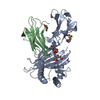
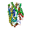
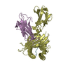
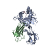
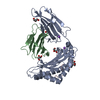
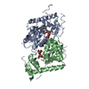
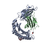
| Title | Crystal structure of PvSUB1 | |||||||||
 Components Components | Subtilisin-like 1 serine protease | |||||||||
 Keywords Keywords |  HYDROLASE / plasmodium egress protease / calcium-binding HYDROLASE / plasmodium egress protease / calcium-binding | |||||||||
| Function / homology |  Function and homology informationsubtilisin / serine-type endopeptidase activity / proteolysis / metal ion binding Function and homology informationsubtilisin / serine-type endopeptidase activity / proteolysis / metal ion bindingSimilarity search - Function | |||||||||
| Biological species |  Plasmodium vivax (malaria parasite P. vivax) Plasmodium vivax (malaria parasite P. vivax) | |||||||||
| Method | X-RAY DIFFRACTION / SYNCHROTRON / MOLECULAR REPLACEMENT / Resolution: 2.7 Å | |||||||||
 Authors Authors | Giganti, D. / Bouillon, A. / Martinez, M. / Weber, P. / Girard-Blanc, C. / Petres, S. / Haouz, A. / Barale, J.C. / Alzari, P.M. | |||||||||
| Funding support |  France, 1items France, 1items
| |||||||||
 Citation Citation | Journal: Nat Commun / Year: 2014 Title: A novel Plasmodium-specific prodomain fold regulates the malaria drug target SUB1 subtilase. Authors: Giganti, D. / Bouillon, A. / Tawk, L. / Robert, F. / Martinez, M. / Crublet, E. / Weber, P. / Girard-Blanc, C. / Petres, S. / Haouz, A. / Hernandez, J.F. / Mercereau-Puijalon, O. / Alzari, P.M. / Barale, J.C. | |||||||||
| History |
|
- Structure visualization
| Structure viewer | Molecule: MolmilJmol/JSmol |
|---|
- Downloads & links
-Download
| PDBx/mmCIF format | 4tr2.cif.gz | 388.4 KB | Display | PDBx/mmCIF format |
|---|---|---|---|---|
| PDB format | pdb4tr2.ent.gz | 316.6 KB | Display | PDB format |
| PDBx/mmJSON format | 4tr2.json.gz | Tree view | PDBx/mmJSON format | |
| Others |  Other downloads Other downloads |
-Validation report
| Arichive directory | https://data.pdbj.org/pub/pdb/validation_reports/tr/4tr2ftp://data.pdbj.org/pub/pdb/validation_reports/tr/4tr2 | HTTPS FTP |
|---|
-Related structure data
| Related structure data | 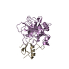 1cgiS 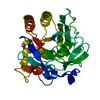 1dbiS 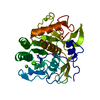 1ea7S  1ic6S  1lw6S  1r0rS  2tecS S: Starting model for refinement |
|---|---|
| Similar structure data |


-Links
 PDBj
PDBj

- Assembly
| Deposited unit | 
| ||||||||
|---|---|---|---|---|---|---|---|---|---|
| 1 | 
| ||||||||
| 2 | 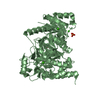
| ||||||||
| 3 |
| ||||||||
| Unit cell |
|
-Components
| #1: Protein | Mass: 73540.398 Da / Num. of mol.: 2 / Fragment: Residues 26-630 Source method: isolated from a genetically manipulated source Source: (gene. exp.) Plasmodium vivax (malaria parasite P. vivax)Gene: sub1 / Production host:   Spodoptera frugiperda (fall armyworm) / References: UniProt: E6Y8B9 Spodoptera frugiperda (fall armyworm) / References: UniProt: E6Y8B9#2: Chemical | ChemComp-CA /   Mass: 40.078 Da / Num. of mol.: 8 / Source method: obtained synthetically / Formula: Ca Mass: 40.078 Da / Num. of mol.: 8 / Source method: obtained synthetically / Formula: Ca#3: Chemical | ChemComp-PO4 / Phosphate  Mass: 94.971 Da / Num. of mol.: 5 / Source method: obtained synthetically / Formula: PO4 Mass: 94.971 Da / Num. of mol.: 5 / Source method: obtained synthetically / Formula: PO4#4: Water | ChemComp-HOH / | Water Mass: 18.015 Da / Num. of mol.: 15 / Source method: isolated from a natural source / Formula: H2O Mass: 18.015 Da / Num. of mol.: 15 / Source method: isolated from a natural source / Formula: H2O |
|---|
-Experimental details
-Experiment
| Experiment | Method: X-RAY DIFFRACTION |
|---|
- Sample preparation
| Crystal | Density Matthews: 2.21 Å3/Da / Density % sol: 44.27 % |
|---|---|
| Crystal grow | Temperature: 291 K / Method: vapor diffusion, hanging drop / pH: 4.2 Details: 1.6 M NaH2PO4, 0.4 M K2HPO4, 0.1 M phosphate citrate |
-Data collection
| Diffraction | Mean temperature: 100 K |
|---|---|
| Diffraction source | Source: SYNCHROTRON / Site: SOLEIL / Beamline: PROXIMA 1 / Wavelength: 0.9801 Å |
| Detector | Type: ADSC QUANTUM 315r / Detector: CCD / Date: Dec 16, 2010 |
| Radiation | Protocol: SINGLE WAVELENGTH / Monochromatic (M) / Laue (L): M / Scattering type: x-ray |
| Radiation wavelength | Wavelength: 0.9801 Å / Relative weight: 1 |
| Reflection | Resolution: 2.7→45 Å / Num. obs: 37100 / % possible obs: 99.4 % / Redundancy: 8.6 % / Biso Wilson estimate: 60.13 Å2 / Rmerge(I) obs: 0.137 / Net I/σ(I): 9.2 |
| Reflection shell | Resolution: 2.7→2.85 Å / Redundancy: 8.8 % / Rmerge(I) obs: 0.726 / Mean I/σ(I) obs: 2.6 / % possible all: 100 |
- Processing
| Software | Name: BUSTER / Version: 2.11.4 / Classification: refinement | ||||||||||||||||||||||||||||||||||||||||||||||||||||||||||||||||||||||||||||||||||||||||||||||||||||||||||||||||||
|---|---|---|---|---|---|---|---|---|---|---|---|---|---|---|---|---|---|---|---|---|---|---|---|---|---|---|---|---|---|---|---|---|---|---|---|---|---|---|---|---|---|---|---|---|---|---|---|---|---|---|---|---|---|---|---|---|---|---|---|---|---|---|---|---|---|---|---|---|---|---|---|---|---|---|---|---|---|---|---|---|---|---|---|---|---|---|---|---|---|---|---|---|---|---|---|---|---|---|---|---|---|---|---|---|---|---|---|---|---|---|---|---|---|---|---|
| Refinement | Method to determine structure: MOLECULAR REPLACEMENT Starting model: PDB entries 2TEC, 1DBI, 1CGI, 1R0R, 1LW6, 1EA7, and 1IC6 Resolution: 2.7→45.1 Å / Cor.coef. Fo:Fc: 0.9288 / Cor.coef. Fo:Fc free: 0.8957 / SU R Cruickshank DPI: 0.537 / Cross valid method: THROUGHOUT / σ(F): 0 / SU R Blow DPI: 0.541 / SU Rfree Blow DPI: 0.29 / SU Rfree Cruickshank DPI: 0.294
| ||||||||||||||||||||||||||||||||||||||||||||||||||||||||||||||||||||||||||||||||||||||||||||||||||||||||||||||||||
| Displacement parameters | Biso mean: 61.98 Å2
| ||||||||||||||||||||||||||||||||||||||||||||||||||||||||||||||||||||||||||||||||||||||||||||||||||||||||||||||||||
| Refine analyze | Luzzati coordinate error obs: 0.418 Å | ||||||||||||||||||||||||||||||||||||||||||||||||||||||||||||||||||||||||||||||||||||||||||||||||||||||||||||||||||
| Refinement step | Cycle: 1 / Resolution: 2.7→45.1 Å
| ||||||||||||||||||||||||||||||||||||||||||||||||||||||||||||||||||||||||||||||||||||||||||||||||||||||||||||||||||
| Refine LS restraints |
| ||||||||||||||||||||||||||||||||||||||||||||||||||||||||||||||||||||||||||||||||||||||||||||||||||||||||||||||||||
| LS refinement shell | Resolution: 2.7→2.77 Å / Total num. of bins used: 19
| ||||||||||||||||||||||||||||||||||||||||||||||||||||||||||||||||||||||||||||||||||||||||||||||||||||||||||||||||||
| Refinement TLS params. | Method: refined / Refine-ID: X-RAY DIFFRACTION
| ||||||||||||||||||||||||||||||||||||||||||||||||||||||||||||||||||||||||||||||||||||||||||||||||||||||||||||||||||
| Refinement TLS group |
|
