 Movie
Movie Controller

[English] 日本語
 Yorodumi
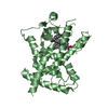
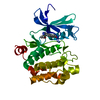
- PDB-6dj6: The X-ray crystal structure of the Streptococcus pneumoniae Fatty... -
+ Open data

- Basic information
| Entry | Database: PDB / ID: 6dj6 | ||||||
|---|---|---|---|---|---|---|---|
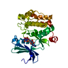
| Title | The X-ray crystal structure of the Streptococcus pneumoniae Fatty Acid Kinase (Fak) B2 protein loaded with cis-oleic acid to 1.9 Angstrom resolution | ||||||
 Components Components | Fatty Acid Kinase (Fak) B2 protein (SPR1019) | ||||||
 Keywords Keywords |  TRANSFERASE / Streptococcus pneumoniae / Fatty acid kinase / oleic acid TRANSFERASE / Streptococcus pneumoniae / Fatty acid kinase / oleic acid | ||||||
| Function / homology |  Function and homology information Function and homology information | ||||||
| Biological species |   Streptococcus pneumoniae (bacteria) Streptococcus pneumoniae (bacteria) | ||||||
| Method | X-RAY DIFFRACTION / SYNCHROTRON / MOLECULAR REPLACEMENT / Resolution: 1.9 Å | ||||||
 Authors Authors | Cuypers, M.G. / Subramanian, C. / White, S.W. / Rock, C.O. | ||||||
| Funding support |  United States, 1items United States, 1items
| ||||||
 Citation Citation | Journal: To Be Published Title: The X-ray crystal structure of the Streptococcus pneumoniae Fatty Acid Kinase (Fak) B2 protein (SPR1019) loaded with cis-oleic acid to 1.9 Angstrom resolution Authors: Cuypers, M.G. / Subramanian, C. / White, S.W. / Rock, C.O. | ||||||
| History |
|
- Structure visualization
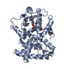
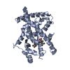
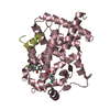
| Structure viewer | Molecule: MolmilJmol/JSmol |
|---|
- Downloads & links
-Download
| PDBx/mmCIF format | 6dj6.cif.gz | 139.6 KB | Display | PDBx/mmCIF format |
|---|---|---|---|---|
| PDB format | pdb6dj6.ent.gz | 105.8 KB | Display | PDB format |
| PDBx/mmJSON format | 6dj6.json.gz | Tree view | PDBx/mmJSON format | |
| Others |  Other downloads Other downloads |
-Validation report
| Arichive directory | https://data.pdbj.org/pub/pdb/validation_reports/dj/6dj6ftp://data.pdbj.org/pub/pdb/validation_reports/dj/6dj6 | HTTPS FTP |
|---|
-Related structure data
| Related structure data | |
|---|---|
| Similar structure data |


-Links
 PDBj
PDBj


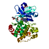
- Assembly
| Deposited unit | 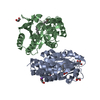
| ||||||||
|---|---|---|---|---|---|---|---|---|---|
| 1 | 
| ||||||||
| 2 | 
| ||||||||
| Unit cell |
|
-Components
| #1: Protein | Mass: 34324.215 Da / Num. of mol.: 2 Source method: isolated from a genetically manipulated source Source: (gene. exp.) Streptococcus pneumoniae (bacteria) / Gene: SPR1019 / Production host: Escherichia coli (E. coli) / References: UniProt: A0A0I9AIE4#2: Chemical | ChemComp-GOL / Glycerol  Mass: 92.094 Da / Num. of mol.: 5 / Source method: obtained synthetically / Formula: C3H8O3 Mass: 92.094 Da / Num. of mol.: 5 / Source method: obtained synthetically / Formula: C3H8O3#3: Chemical | Oleic acid  Mass: 282.461 Da / Num. of mol.: 2 / Source method: obtained synthetically / Formula: C18H34O2 Mass: 282.461 Da / Num. of mol.: 2 / Source method: obtained synthetically / Formula: C18H34O2#4: Chemical | ChemComp-NA / |   Mass: 22.990 Da / Num. of mol.: 1 / Source method: obtained synthetically / Formula: Na Mass: 22.990 Da / Num. of mol.: 1 / Source method: obtained synthetically / Formula: Na#5: Water | ChemComp-HOH / | Water Mass: 18.015 Da / Num. of mol.: 551 / Source method: isolated from a natural source / Formula: H2O Mass: 18.015 Da / Num. of mol.: 551 / Source method: isolated from a natural source / Formula: H2O |
|---|
-Experimental details
-Experiment
| Experiment | Method: X-RAY DIFFRACTION / Number of used crystals: 1 |
|---|
- Sample preparation
| Crystal | Density Matthews: 2.45 Å3/Da / Density % sol: 49.75 % |
|---|---|
| Crystal grow | Temperature: 293 K / Method: vapor diffusion, hanging drop / pH: 7.4 Details: 0.1M HEPES pH 7.4, 10% v/v isopropanol, 20% w/v PEG4000 Temp details: controlled temperature room |
-Data collection
| Diffraction | Mean temperature: 100 K / Ambient temp details: pre frozen in Liquid N2 |
|---|---|
| Diffraction source | Source: SYNCHROTRON / Site: APS / Beamline: 22-ID / Wavelength: 1 Å |
| Detector | Type: DECTRIS EIGER X 16M / Detector: PIXEL / Date: Mar 15, 2018 |
| Radiation | Protocol: SINGLE WAVELENGTH / Monochromatic (M) / Laue (L): M / Scattering type: x-ray |
| Radiation wavelength | Wavelength: 1 Å / Relative weight: 1 |
| Reflection | Resolution: 1.9→74.92 Å / Num. obs: 52631 / % possible obs: 98.3 % / Redundancy: 3.7 % / Biso Wilson estimate: 17.034 Å2 / CC1/2: 0.993 / Rmerge(I) obs: 0.109 / Rpim(I) all: 0.067 / Rrim(I) all: 0.129 / Χ2: 0.95 / Net I/σ(I): 9.4 |
| Reflection shell | Resolution: 1.9→1.94 Å / Redundancy: 3.7 % / Rmerge(I) obs: 0.683 / Mean I/σ(I) obs: 1.9 / Num. unique obs: 3366 / CC1/2: 0.479 / Rpim(I) all: 0.51 / Rrim(I) all: 0.896 / Χ2: 1.07 / % possible all: 99.6 |
- Processing
| Software |
| ||||||||||||||||||||||||||||||||||||||||||||||||||||||||||||||||||||||||||||||||||||||||||||||||||||||||||||||||||||||||||||||||||||||||||||||||||||||||||||||||||||||||||||||||||||||
|---|---|---|---|---|---|---|---|---|---|---|---|---|---|---|---|---|---|---|---|---|---|---|---|---|---|---|---|---|---|---|---|---|---|---|---|---|---|---|---|---|---|---|---|---|---|---|---|---|---|---|---|---|---|---|---|---|---|---|---|---|---|---|---|---|---|---|---|---|---|---|---|---|---|---|---|---|---|---|---|---|---|---|---|---|---|---|---|---|---|---|---|---|---|---|---|---|---|---|---|---|---|---|---|---|---|---|---|---|---|---|---|---|---|---|---|---|---|---|---|---|---|---|---|---|---|---|---|---|---|---|---|---|---|---|---|---|---|---|---|---|---|---|---|---|---|---|---|---|---|---|---|---|---|---|---|---|---|---|---|---|---|---|---|---|---|---|---|---|---|---|---|---|---|---|---|---|---|---|---|---|---|---|---|
| Refinement | Method to determine structure: MOLECULAR REPLACEMENT Starting model: MODBASE sequence based generated 3D model Resolution: 1.9→54.15 Å / Cor.coef. Fo:Fc: 0.964 / Cor.coef. Fo:Fc free: 0.931 / SU B: 4.349 / SU ML: 0.119 / Cross valid method: THROUGHOUT / ESU R: 0.136 / ESU R Free: 0.14 / Details: HYDROGENS HAVE BEEN ADDED IN THE RIDING POSITIONS
| ||||||||||||||||||||||||||||||||||||||||||||||||||||||||||||||||||||||||||||||||||||||||||||||||||||||||||||||||||||||||||||||||||||||||||||||||||||||||||||||||||||||||||||||||||||||
| Solvent computation | Ion probe radii: 0.8 Å / Shrinkage radii: 0.8 Å / VDW probe radii: 1.2 Å | ||||||||||||||||||||||||||||||||||||||||||||||||||||||||||||||||||||||||||||||||||||||||||||||||||||||||||||||||||||||||||||||||||||||||||||||||||||||||||||||||||||||||||||||||||||||
| Displacement parameters | Biso mean: 24.118 Å2
| ||||||||||||||||||||||||||||||||||||||||||||||||||||||||||||||||||||||||||||||||||||||||||||||||||||||||||||||||||||||||||||||||||||||||||||||||||||||||||||||||||||||||||||||||||||||
| Refinement step | Cycle: 1 / Resolution: 1.9→54.15 Å
| ||||||||||||||||||||||||||||||||||||||||||||||||||||||||||||||||||||||||||||||||||||||||||||||||||||||||||||||||||||||||||||||||||||||||||||||||||||||||||||||||||||||||||||||||||||||
| Refine LS restraints |
|