 Movie
Movie Controller

[English] 日本語
 Yorodumi
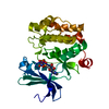
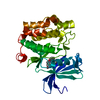
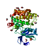
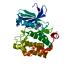
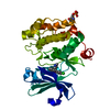
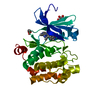
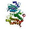
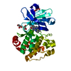
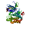
- PDB-6ayd: Pim1 complexed with N-(6-(4-hydroxyphenyl)-1H-indazol-3-yl)cyclop... -
+ Open data

- Basic information
| Entry | Database: PDB / ID: 6ayd | ||||||
|---|---|---|---|---|---|---|---|
| Title | Pim1 complexed with N-(6-(4-hydroxyphenyl)-1H-indazol-3-yl)cyclopropanecarboxamide | ||||||
 Components Components | Serine/threonine-protein kinase pim-1 | ||||||
 Keywords Keywords | Transferase/Transferase Inhibitor /  Kinase / Transferase-Transferase Inhibitor complex Kinase / Transferase-Transferase Inhibitor complex | ||||||
| Function / homology |  Function and homology information Function and homology informationpositive regulation of cardioblast proliferation / cellular detoxification / regulation of hematopoietic stem cell proliferation / vitamin D receptor signaling pathway / STAT5 activation downstream of FLT3 ITD mutants / transcription factor binding / ribosomal small subunit binding / positive regulation of cyclin-dependent protein serine/threonine kinase activity / positive regulation of cardiac muscle cell proliferation / positive regulation of TORC1 signaling ...positive regulation of cardioblast proliferation / cellular detoxification / regulation of hematopoietic stem cell proliferation / vitamin D receptor signaling pathway / STAT5 activation downstream of FLT3 ITD mutants / transcription factor binding / ribosomal small subunit binding / positive regulation of cyclin-dependent protein serine/threonine kinase activity / positive regulation of cardiac muscle cell proliferation / positive regulation of TORC1 signaling / Signaling by FLT3 fusion proteins / negative regulation of innate immune response / positive regulation of brown fat cell differentiation / protein serine/threonine kinase activator activity / regulation of transmembrane transporter activity / positive regulation of protein serine/threonine kinase activity / negative regulation of DNA-binding transcription factor activity / cellular response to type II interferon / manganese ion binding / Interleukin-4 and Interleukin-13 signaling / protein autophosphorylation / protein stabilization / non-specific serine/threonine protein kinase / cell cycle / protein phosphorylation / protein serine kinase activity / protein serine/threonine kinase activity / apoptotic process / nucleolus / negative regulation of apoptotic process / positive regulation of DNA-templated transcription / nucleoplasm / ATP binding / nucleus / plasma membrane / cytosol / cytoplasmSimilarity search - Function | ||||||
| Biological species |  Homo sapiens (human) Homo sapiens (human) | ||||||
| Method | X-RAY DIFFRACTION / MOLECULAR REPLACEMENT / Resolution: 3 Å | ||||||
 Authors Authors | Shewchuk, L.M. / Henley, Z.A. | ||||||
 Citation Citation | Journal: ACS Med Chem Lett / Year: 2017 Title: From PIM1 to PI3K delta via GSK3 beta : Target Hopping through the Kinome. Authors: Henley, Z.A. / Bax, B.D. / Inglesby, L.M. / Champigny, A. / Gaines, S. / Faulder, P. / Le, J. / Thomas, D.A. / Washio, Y. / Baldwin, I.R. | ||||||
| History |
|
- Structure visualization
| Structure viewer | Molecule: MolmilJmol/JSmol |
|---|
- Downloads & links
-Download
| PDBx/mmCIF format | 6ayd.cif.gz | 127.6 KB | Display | PDBx/mmCIF format |
|---|---|---|---|---|
| PDB format | pdb6ayd.ent.gz | 97.8 KB | Display | PDB format |
| PDBx/mmJSON format | 6ayd.json.gz | Tree view | PDBx/mmJSON format | |
| Others |  Other downloads Other downloads |
-Validation report
| Arichive directory | https://data.pdbj.org/pub/pdb/validation_reports/ay/6aydftp://data.pdbj.org/pub/pdb/validation_reports/ay/6ayd | HTTPS FTP |
|---|
-Related structure data
| Related structure data |  5oy4C  2bikS S: Starting model for refinement C: citing same article ( |
|---|---|
| Similar structure data |


-Links
 PDBj
PDBj

- Assembly
| Deposited unit | 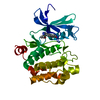
| ||||||||
|---|---|---|---|---|---|---|---|---|---|
| 1 |
| ||||||||
| Unit cell |
|
-Components
| #1: Protein | Mass: 38062.148 Da / Num. of mol.: 1 / Fragment: UNP residues 1-312 / Mutation: R250G Source method: isolated from a genetically manipulated source Source: (gene. exp.) Homo sapiens (human) / Gene: PIM1 / Production host:  Escherichia coli (E. coli) Escherichia coli (E. coli)References: UniProt: P11309, non-specific serine/threonine protein kinase |
|---|---|
| #2: Chemical | ChemComp-C2J /   Mass: 293.320 Da / Num. of mol.: 1 / Source method: obtained synthetically / Formula: C17H15N3O2 Mass: 293.320 Da / Num. of mol.: 1 / Source method: obtained synthetically / Formula: C17H15N3O2 |
| #3: Water | ChemComp-HOH / Water Mass: 18.015 Da / Num. of mol.: 34 / Source method: isolated from a natural source / Formula: H2O Mass: 18.015 Da / Num. of mol.: 34 / Source method: isolated from a natural source / Formula: H2O |
-Experimental details
-Experiment
| Experiment | Method: X-RAY DIFFRACTION / Number of used crystals: 1 |
|---|
- Sample preparation
| Crystal | Density Matthews: 2.88 Å3/Da / Density % sol: 57.29 % |
|---|---|
| Crystal grow | Temperature: 277 K / Method: vapor diffusion Details: 1M succinate pH 7.0, 0.1M BisTrisPropane, 1% 1,6-hexanediol |
-Data collection
| Diffraction | Mean temperature: 93 K |
|---|---|
| Diffraction source | Source: ROTATING ANODE / Type: RIGAKU MICROMAX-007 HF / Wavelength: 1.54 Å |
| Detector | Type: MARRESEARCH / Detector: CCD / Date: Aug 9, 2006 |
| Radiation | Protocol: SINGLE WAVELENGTH / Monochromatic (M) / Laue (L): M / Scattering type: x-ray |
| Radiation wavelength | Wavelength: 1.54 Å / Relative weight: 1 |
| Reflection | Resolution: 3→20 Å / Num. obs: 7834 / % possible obs: 94 % / Redundancy: 5.5 % / Rmerge(I) obs: 0.13 / Net I/σ(I): 15 |
| Reflection shell | Resolution: 3→3.078 Å / Rmerge(I) obs: 0.54 / Num. unique obs: 385 / % possible all: 63 |
- Processing
| Software |
| ||||||||||||||||||||||||||||||||||||||||||||||||||||||||||||
|---|---|---|---|---|---|---|---|---|---|---|---|---|---|---|---|---|---|---|---|---|---|---|---|---|---|---|---|---|---|---|---|---|---|---|---|---|---|---|---|---|---|---|---|---|---|---|---|---|---|---|---|---|---|---|---|---|---|---|---|---|---|
| Refinement | Method to determine structure: MOLECULAR REPLACEMENT Starting model: 2BIK Resolution: 3→20 Å / Cor.coef. Fo:Fc: 0.949 / Cor.coef. Fo:Fc free: 0.885 / SU B: 36.658 / SU ML: 0.314 / Cross valid method: THROUGHOUT / σ(F): 0 / ESU R Free: 0.383 Details: HYDROGENS HAVE BEEN ADDED IN THE RIDING POSITIONS U VALUES : WITH TLS ADDED
| ||||||||||||||||||||||||||||||||||||||||||||||||||||||||||||
| Solvent computation | Ion probe radii: 0.8 Å / Shrinkage radii: 0.8 Å / VDW probe radii: 1.2 Å | ||||||||||||||||||||||||||||||||||||||||||||||||||||||||||||
| Displacement parameters | Biso max: 134.48 Å2 / Biso mean: 73.88 Å2 / Biso min: 23.83 Å2
| ||||||||||||||||||||||||||||||||||||||||||||||||||||||||||||
| Refinement step | Cycle: final / Resolution: 3→20 Å
| ||||||||||||||||||||||||||||||||||||||||||||||||||||||||||||
| Refine LS restraints |
| ||||||||||||||||||||||||||||||||||||||||||||||||||||||||||||
| LS refinement shell | Resolution: 3→3.078 Å / Rfactor Rfree error: 0 / Total num. of bins used: 20
| ||||||||||||||||||||||||||||||||||||||||||||||||||||||||||||
| Refinement TLS params. | Method: refined / Origin x: -8.681 Å / Origin y: -40.228 Å / Origin z: -0.63 Å
|
