 Movie
Movie Controller

[English] 日本語
 Yorodumi
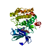
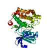
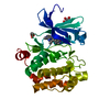
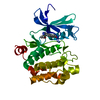
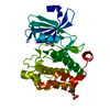
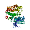
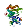
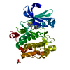
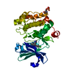
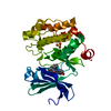
- PDB-5kge: Crystal structure of PIM1 with inhibitor: 5-(3,4-dichlorophenyl)-... -
+ Open data

- Basic information
| Entry | Database: PDB / ID: 5kge | ||||||
|---|---|---|---|---|---|---|---|
| Title | Crystal structure of PIM1 with inhibitor: 5-(3,4-dichlorophenyl)-1~{H}-pyrazol-3-amine | ||||||
 Components Components | Serine/threonine-protein kinase pim-1 | ||||||
 Keywords Keywords | TRANSFERASE/TRANSFERASE INHIBITOR /  Kinase / transferase / Serine/threonine-protein kinase pim-1 / TRANSFERASE-TRANSFERASE INHIBITOR complex Kinase / transferase / Serine/threonine-protein kinase pim-1 / TRANSFERASE-TRANSFERASE INHIBITOR complex | ||||||
| Function / homology |  Function and homology information Function and homology informationpositive regulation of cardioblast proliferation / cellular detoxification / regulation of hematopoietic stem cell proliferation / vitamin D receptor signaling pathway / STAT5 activation downstream of FLT3 ITD mutants / transcription factor binding / ribosomal small subunit binding / positive regulation of cyclin-dependent protein serine/threonine kinase activity / positive regulation of cardiac muscle cell proliferation / positive regulation of TORC1 signaling ...positive regulation of cardioblast proliferation / cellular detoxification / regulation of hematopoietic stem cell proliferation / vitamin D receptor signaling pathway / STAT5 activation downstream of FLT3 ITD mutants / transcription factor binding / ribosomal small subunit binding / positive regulation of cyclin-dependent protein serine/threonine kinase activity / positive regulation of cardiac muscle cell proliferation / positive regulation of TORC1 signaling / Signaling by FLT3 fusion proteins / negative regulation of innate immune response / positive regulation of brown fat cell differentiation / protein serine/threonine kinase activator activity / regulation of transmembrane transporter activity / positive regulation of protein serine/threonine kinase activity / negative regulation of DNA-binding transcription factor activity / cellular response to type II interferon / manganese ion binding / Interleukin-4 and Interleukin-13 signaling / protein autophosphorylation / protein stabilization / non-specific serine/threonine protein kinase / cell cycle / protein phosphorylation / protein serine kinase activity / protein serine/threonine kinase activity / apoptotic process / nucleolus / negative regulation of apoptotic process / positive regulation of DNA-templated transcription / nucleoplasm / ATP binding / nucleus / plasma membrane / cytosol / cytoplasmSimilarity search - Function | ||||||
| Biological species |  Homo sapiens (human) Homo sapiens (human) | ||||||
| Method | X-RAY DIFFRACTION / SYNCHROTRON / MOLECULAR REPLACEMENT / Resolution: 2.23 Å | ||||||
 Authors Authors | Ferguson, A.D. | ||||||
 Citation Citation | Journal: To Be Published Title: Structure of PIM1 in complex with inhibitor Authors: Ferguson, A.D. | ||||||
| History |
|
- Structure visualization
| Structure viewer | Molecule: MolmilJmol/JSmol |
|---|
- Downloads & links
-Download
| PDBx/mmCIF format | 5kge.cif.gz | 130.9 KB | Display | PDBx/mmCIF format |
|---|---|---|---|---|
| PDB format | pdb5kge.ent.gz | 100.7 KB | Display | PDB format |
| PDBx/mmJSON format | 5kge.json.gz | Tree view | PDBx/mmJSON format | |
| Others |  Other downloads Other downloads |
-Validation report
| Arichive directory | https://data.pdbj.org/pub/pdb/validation_reports/kg/5kgeftp://data.pdbj.org/pub/pdb/validation_reports/kg/5kge | HTTPS FTP |
|---|
-Related structure data
| Related structure data |  5kgdC  5kggC  5kgiC  5kgkC  4dtkS C: citing same article ( S: Starting model for refinement |
|---|---|
| Similar structure data |


-Links
 PDBj
PDBj

- Assembly
| Deposited unit | 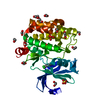
| ||||||||
|---|---|---|---|---|---|---|---|---|---|
| 1 |
| ||||||||
| Unit cell |
|
-Components
| #1: Protein | Mass: 31905.203 Da / Num. of mol.: 1 / Fragment: UNP residues 30-305 Source method: isolated from a genetically manipulated source Source: (gene. exp.) Homo sapiens (human) / Gene: PIM1 / Production host:  Escherichia coli (E. coli) Escherichia coli (E. coli)References: UniProt: P11309, non-specific serine/threonine protein kinase | ||||||
|---|---|---|---|---|---|---|---|
| #2: Chemical | ChemComp-EDO / Ethylene glycol  Mass: 62.068 Da / Num. of mol.: 15 / Source method: obtained synthetically / Formula: C2H6O2 Mass: 62.068 Da / Num. of mol.: 15 / Source method: obtained synthetically / Formula: C2H6O2#3: Chemical | ChemComp-6SN / |   Mass: 228.078 Da / Num. of mol.: 1 / Source method: obtained synthetically / Formula: C9H7Cl2N3 Mass: 228.078 Da / Num. of mol.: 1 / Source method: obtained synthetically / Formula: C9H7Cl2N3#4: Chemical | Sulfate  Mass: 96.063 Da / Num. of mol.: 2 / Source method: obtained synthetically / Formula: SO4 Mass: 96.063 Da / Num. of mol.: 2 / Source method: obtained synthetically / Formula: SO4#5: Water | ChemComp-HOH / | Water Mass: 18.015 Da / Num. of mol.: 56 / Source method: isolated from a natural source / Formula: H2O Mass: 18.015 Da / Num. of mol.: 56 / Source method: isolated from a natural source / Formula: H2O |
-Experimental details
-Experiment
| Experiment | Method: X-RAY DIFFRACTION / Number of used crystals: 1 |
|---|
- Sample preparation
| Crystal | Density Matthews: 3.43 Å3/Da / Density % sol: 64.13 % |
|---|---|
| Crystal grow | Temperature: 293 K / Method: vapor diffusion, hanging drop / pH: 7.3 Details: 18% PEG 3350, 0.2 M Na2SO4, 6% ethylene glycol, 0.1 M Tris, pH 7.3 |
-Data collection
| Diffraction | Mean temperature: 100 K |
|---|---|
| Diffraction source | Source: SYNCHROTRON / Site: APS  / Beamline: 17-ID / Wavelength: 1 Å / Beamline: 17-ID / Wavelength: 1 Å |
| Detector | Type: DECTRIS PILATUS 6M / Detector: PIXEL / Date: Jun 20, 2011 |
| Radiation | Protocol: SINGLE WAVELENGTH / Monochromatic (M) / Laue (L): M / Scattering type: x-ray |
| Radiation wavelength | Wavelength: 1 Å / Relative weight: 1 |
| Reflection | Resolution: 2.23→58.19 Å / Num. obs: 21136 / % possible obs: 99.7 % / Redundancy: 11.3 % / Biso Wilson estimate: 49.74 Å2 / Rmerge(I) obs: 0.067 / Net I/σ(I): 24.4 |
| Reflection shell | Resolution: 2.23→2.35 Å / Redundancy: 11.4 % / Rmerge(I) obs: 0.571 / Mean I/σ(I) obs: 4.7 / % possible all: 100 |
- Processing
| Software |
| ||||||||||||||||||||||||||||||||||||||||||||||||||||||||||||||||||||||||||||||||||||||||||||||||||||||||||||||||||
|---|---|---|---|---|---|---|---|---|---|---|---|---|---|---|---|---|---|---|---|---|---|---|---|---|---|---|---|---|---|---|---|---|---|---|---|---|---|---|---|---|---|---|---|---|---|---|---|---|---|---|---|---|---|---|---|---|---|---|---|---|---|---|---|---|---|---|---|---|---|---|---|---|---|---|---|---|---|---|---|---|---|---|---|---|---|---|---|---|---|---|---|---|---|---|---|---|---|---|---|---|---|---|---|---|---|---|---|---|---|---|---|---|---|---|---|
| Refinement | Method to determine structure: MOLECULAR REPLACEMENT Starting model: 4DTK Resolution: 2.23→58.19 Å / Cor.coef. Fo:Fc: 0.928 / Cor.coef. Fo:Fc free: 0.926 / Rfactor Rfree error: 0.01 / SU R Cruickshank DPI: 0.198 / Cross valid method: THROUGHOUT / σ(F): 0 / SU R Blow DPI: 0.19 / SU Rfree Blow DPI: 0.151 / SU Rfree Cruickshank DPI: 0.155
| ||||||||||||||||||||||||||||||||||||||||||||||||||||||||||||||||||||||||||||||||||||||||||||||||||||||||||||||||||
| Displacement parameters | Biso mean: 53.58 Å2
| ||||||||||||||||||||||||||||||||||||||||||||||||||||||||||||||||||||||||||||||||||||||||||||||||||||||||||||||||||
| Refine analyze | Luzzati coordinate error obs: 0.3 Å | ||||||||||||||||||||||||||||||||||||||||||||||||||||||||||||||||||||||||||||||||||||||||||||||||||||||||||||||||||
| Refinement step | Cycle: 1 / Resolution: 2.23→58.19 Å
| ||||||||||||||||||||||||||||||||||||||||||||||||||||||||||||||||||||||||||||||||||||||||||||||||||||||||||||||||||
| Refine LS restraints |
| ||||||||||||||||||||||||||||||||||||||||||||||||||||||||||||||||||||||||||||||||||||||||||||||||||||||||||||||||||
| LS refinement shell | Resolution: 2.23→2.34 Å / Rfactor Rfree error: 0 / Total num. of bins used: 11
| ||||||||||||||||||||||||||||||||||||||||||||||||||||||||||||||||||||||||||||||||||||||||||||||||||||||||||||||||||
| Refinement TLS params. | Method: refined / Origin x: 38.8262 Å / Origin y: -12.6165 Å / Origin z: 0.3404 Å
| ||||||||||||||||||||||||||||||||||||||||||||||||||||||||||||||||||||||||||||||||||||||||||||||||||||||||||||||||||
| Refinement TLS group | Selection details: { A|* } |
