[English] 日本語
Yorodumi
- PDB-6cng: The X-ray crystal structure of the Streptococcus pneumoniae Fatty... -

+
Open data


ID or keywords:

Loading...

-
Basic information

Entry
Database: PDB / ID: 6cng
TitleThe X-ray crystal structure of the Streptococcus pneumoniae Fatty Acid Kinase (Fak) B3 protein loaded with linoleic acid to 1.47 Angstrom resolution
ComponentsFatty Acid Kinase (Fak) B3 protein
KeywordsTRANSFERASE / Streptococcus pneumoniae / Fatty acid kinase / Linoleic acid
Function / homology
Function and homology information


DegV, N-terminal domain, peripheral subdomain / Hypothetical Protein Tm841; Chain: A;domain 3 - #10 / Dihydroxyacetone kinase; domain 1 / DegV / DegV, C-terminal domain / Uncharacterised protein, DegV family COG1307 / DegV domain profile. / Hypothetical Protein Tm841; Chain: A;domain 3 / Rubrerythrin, domain 2 / Single Sheet ...DegV, N-terminal domain, peripheral subdomain / Hypothetical Protein Tm841; Chain: A;domain 3 - #10 / Dihydroxyacetone kinase; domain 1 / DegV / DegV, C-terminal domain / Uncharacterised protein, DegV family COG1307 / DegV domain profile. / Hypothetical Protein Tm841; Chain: A;domain 3 / Rubrerythrin, domain 2 / Single Sheet / Rossmann fold / 2-Layer Sandwich / 3-Layer(aba) Sandwich / Mainly Beta / Alpha Beta
Similarity search - Domain/homology
ACETATE ION / LINOLEIC ACID / DegV domain-containing protein spr0652
Similarity search - Component
Biological speciesStreptococcus pneumoniae (bacteria)
MethodX-RAY DIFFRACTION / SYNCHROTRON / MOLECULAR REPLACEMENT / Resolution: 1.47 Å
AuthorsCuypers, M.G. / Subramanian, C. / White, S.W. / Rock, C.O.
Funding support United States, 1items
OrganizationGrant numberCountry
National Institutes of Health/National Institute of General Medical Sciences (NIH/NIGMS) United States
CitationJournal: To Be Published
Title: The X-ray crystal structure of the Streptococcus pneumoniae Fatty Acid Kinase (Fak) B3 protein loaded with linoleic acid to 1.47 Angstrom resolution
Authors: Cuypers, M.G. / Subramanian, C. / White, S.W. / Rock, C.O.
History
DepositionMar 8, 2018Deposition site: RCSB / Processing site: RCSB
Revision 1.0Jul 3, 2019Provider: repository / Type: Initial release
Revision 1.1Dec 4, 2019Group: Author supporting evidence / Category: pdbx_audit_support / Item: _pdbx_audit_support.funding_organization
Revision 1.2Oct 4, 2023Group: Data collection / Database references ...Data collection / Database references / Derived calculations / Refinement description
Category: chem_comp_atom / chem_comp_bond ...chem_comp_atom / chem_comp_bond / database_2 / pdbx_initial_refinement_model / pdbx_struct_conn_angle / struct_conn
Item: _database_2.pdbx_DOI / _database_2.pdbx_database_accession ..._database_2.pdbx_DOI / _database_2.pdbx_database_accession / _pdbx_struct_conn_angle.ptnr1_auth_seq_id / _pdbx_struct_conn_angle.ptnr3_auth_seq_id / _pdbx_struct_conn_angle.value / _struct_conn.pdbx_dist_value / _struct_conn.ptnr1_auth_asym_id / _struct_conn.ptnr1_auth_comp_id / _struct_conn.ptnr1_auth_seq_id / _struct_conn.ptnr1_label_asym_id / _struct_conn.ptnr1_label_atom_id / _struct_conn.ptnr1_label_comp_id / _struct_conn.ptnr1_label_seq_id / _struct_conn.ptnr2_auth_asym_id / _struct_conn.ptnr2_auth_comp_id / _struct_conn.ptnr2_auth_seq_id / _struct_conn.ptnr2_label_asym_id / _struct_conn.ptnr2_label_atom_id / _struct_conn.ptnr2_label_comp_id

-
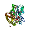
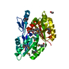
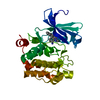
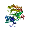
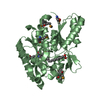
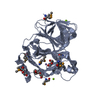
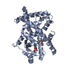
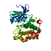
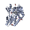
Structure visualization

Structure viewerMolecule:
MolmilJmol/JSmol

Downloads & links

-
Assembly

Deposited unit
A: Fatty Acid Kinase (Fak) B3 protein
D: Fatty Acid Kinase (Fak) B3 protein
hetero molecules


Theoretical massNumber of molelcules
Total (without water)67,83224
Polymers65,7332
Non-polymers2,09922
Water14,322795
1
A: Fatty Acid Kinase (Fak) B3 protein
hetero molecules


Theoretical massNumber of molelcules
Total (without water)33,6569
Polymers32,8661
Non-polymers7908
Water181
TypeNameSymmetry operationNumber
identity operation1_555x,y,z1
MethodPISA
2
D: Fatty Acid Kinase (Fak) B3 protein
hetero molecules


Theoretical massNumber of molelcules
Total (without water)34,17615
Polymers32,8661
Non-polymers1,30914
Water181
TypeNameSymmetry operationNumber
identity operation1_555x,y,z1
MethodPISA
Unit cell
Length a, b, c (Å)52.775, 103.414, 67.469
Angle α, β, γ (deg.)90.00, 112.99, 90.00
Int Tables number4
Space group name H-MP1211

-
Components

-
Protein , 1 types, 2 molecules AD

#1: Protein Fatty Acid Kinase (Fak) B3 protein


Mass: 32866.352 Da / Num. of mol.: 2
Source method: isolated from a genetically manipulated source
Source: (gene. exp.) Streptococcus pneumoniae (strain ATCC BAA-255 / R6) (bacteria)
Strain: ATCC BAA-255 / R6 / Gene: spr0652 / Production host: Escherichia coli (E. coli) / References: UniProt: Q8DQI6

-
Non-polymers , 5 types, 817 molecules

#2: Chemical ChemComp-EIC / LINOLEIC ACID / 9,12-LINOLEIC ACID / Linoleic acid


Mass: 280.445 Da / Num. of mol.: 2 / Source method: obtained synthetically / Formula: C18H32O2
#3: Chemical ChemComp-NA / SODIUM ION


Mass: 22.990 Da / Num. of mol.: 2 / Source method: obtained synthetically / Formula: Na
#4: Chemical
ChemComp-GOL / GLYCEROL / GLYCERIN / PROPANE-1,2,3-TRIOL / Glycerol


Mass: 92.094 Da / Num. of mol.: 13 / Source method: obtained synthetically / Formula: C3H8O3
#5: Chemical
ChemComp-ACT / ACETATE ION / Acetate


Mass: 59.044 Da / Num. of mol.: 5 / Source method: obtained synthetically / Formula: C2H3O2
#6: Water ChemComp-HOH / water / Water


Mass: 18.015 Da / Num. of mol.: 795 / Source method: isolated from a natural source / Formula: H2O

-
Experimental details

-
Experiment

ExperimentMethod: X-RAY DIFFRACTION / Number of used crystals: 1

-
Sample preparation

CrystalDensity Matthews: 2.58 Å3/Da / Density % sol: 52.4 % / Description: prismatic
Crystal growTemperature: 291 K / Method: vapor diffusion, sitting drop / pH: 4.6
Details: 0.1M Na acetate pH 4.6, 8% PEG 4000. Cryofreezing solution soak in motherliquor with 25% glycerol
Temp details: controlled temperature room

-
Data collection

DiffractionMean temperature: 100 K
Ambient temp details: on cryostream (from Liq N2 flashfreeze)
Diffraction sourceSource: SYNCHROTRON / Site: APS / Beamline: 22-ID / Wavelength: 1 Å
DetectorType: RAYONIX MX300-HS / Detector: CCD / Date: Oct 15, 2017
RadiationProtocol: SINGLE WAVELENGTH / Monochromatic (M) / Laue (L): M / Scattering type: x-ray
Radiation wavelengthWavelength: 1 Å / Relative weight: 1
ReflectionResolution: 1.47→62.11 Å / Num. obs: 108472 / % possible obs: 96.2 % / Redundancy: 3 % / Biso Wilson estimate: 17.283 Å2 / CC1/2: 0.999 / Rmerge(I) obs: 0.036 / Rpim(I) all: 0.025 / Rrim(I) all: 0.044 / Net I/σ(I): 15
Reflection shellResolution: 1.47→1.5 Å / Redundancy: 2.9 % / Rmerge(I) obs: 0.544 / Num. unique obs: 5188 / CC1/2: 0.574 / Rpim(I) all: 0.386 / Rrim(I) all: 0.669 / % possible all: 94.1

-
Processing

Software
NameVersionClassification
PHENIX(1.13_2998: ???)refinement
XDSdata reduction
Aimlessdata scaling
PHASERphasing
RefinementMethod to determine structure: MOLECULAR REPLACEMENT
Starting model: 3fdj (modified with MODBASE server)
Resolution: 1.47→43.972 Å / Cross valid method: FREE R-VALUE / σ(F): 6.03 / Phase error: 19.34 / Details: H atoms are isotropic, not H atoms are anisotropic
RfactorNum. reflection% reflection
Rfree0.1647 5335 5.11 %
Rwork0.1365 --
obs0.1521 108436 96.07 %
Solvent computationShrinkage radii: 0.9 Å / VDW probe radii: 1.11 Å
Refinement stepCycle: LAST / Resolution: 1.47→43.972 Å
ProteinNucleic acidLigandSolventTotal
Num. atoms4298 0 140 795 5233
Refine LS restraints
Refine-IDTypeDev idealNumber
X-RAY DIFFRACTIONf_bond_d0.0144496
X-RAY DIFFRACTIONf_angle_d1.1346048
X-RAY DIFFRACTIONf_dihedral_angle_d22.9871683
X-RAY DIFFRACTIONf_chiral_restr0.08702
X-RAY DIFFRACTIONf_plane_restr0.006778
LS refinement shell
Resolution (Å)Rfactor RfreeNum. reflection RfreeRfactor RworkNum. reflection RworkRefine-ID% reflection obs (%)
1.47-1.49540.27972330.25814995X-RAY DIFFRACTION90
1.4954-1.52250.22482760.23695006X-RAY DIFFRACTION89
1.5225-1.55180.23032480.22855037X-RAY DIFFRACTION89
1.5518-1.58350.23522560.23195035X-RAY DIFFRACTION90
1.5835-1.61790.20652540.22025036X-RAY DIFFRACTION90
1.6179-1.65550.24172510.21525107X-RAY DIFFRACTION90
1.6555-1.69690.21722690.21055124X-RAY DIFFRACTION91
1.6969-1.74270.22272860.20445060X-RAY DIFFRACTION90
1.7427-1.79390.20342840.20315110X-RAY DIFFRACTION91
1.7939-1.85180.22462780.19885137X-RAY DIFFRACTION91
1.8518-1.91790.20912670.18635171X-RAY DIFFRACTION92
1.9179-1.99460.19172690.18015160X-RAY DIFFRACTION92
1.9946-2.08520.18792830.17475152X-RAY DIFFRACTION92
2.0852-2.1950.20242600.16335240X-RAY DIFFRACTION92
2.195-2.33220.18232590.15295238X-RAY DIFFRACTION93
2.3322-2.51180.18572520.14665258X-RAY DIFFRACTION93
2.5118-2.76370.16322630.13925244X-RAY DIFFRACTION93
2.7637-3.16170.1752730.12895272X-RAY DIFFRACTION93
3.1617-3.97590.15152880.11235321X-RAY DIFFRACTION93
3.9759-17.70430.15112870.10665337X-RAY DIFFRACTION93
Refinement TLS params.Method: refined / Origin x: 4.88 Å / Origin y: -6.4633 Å / Origin z: -0.3321 Å
111213212223313233
T0.0964 Å20.0035 Å2-0.0062 Å2-0.1312 Å20.007 Å2--0.131 Å2
L0.1819 °20.0072 °2-0.0185 °2-0.0964 °20.1618 °2--0.4733 °2
S-0.0084 Å °0.0003 Å °-0.0017 Å °0.001 Å °0.0057 Å °-0.0116 Å °0.0026 Å °-0.0046 Å °0.0051 Å °
Refinement TLS groupSelection details: all

+
About Yorodumi

-
News

-
Feb 9, 2022. New format data for meta-information of EMDB entries

New format data for meta-information of EMDB entries

  • Version 3 of the EMDB header file is now the official format.
  • The previous official version 1.9 will be removed from the archive.

Related info.:EMDB header

External links:wwPDB to switch to version 3 of the EMDB data model

-
Aug 12, 2020. Covid-19 info

Covid-19 info

URL: https://pdbj.org/emnavi/covid19.php

New page: Covid-19 featured information page in EM Navigator.

Related info.:Covid-19 info / Mar 5, 2020. Novel coronavirus structure data

+
Mar 5, 2020. Novel coronavirus structure data

Novel coronavirus structure data

Related info.:Yorodumi Speices / Aug 12, 2020. Covid-19 info

External links:COVID-19 featured content - PDBj / Molecule of the Month (242):Coronavirus Proteases

+
Jan 31, 2019. EMDB accession codes are about to change! (news from PDBe EMDB page)

EMDB accession codes are about to change! (news from PDBe EMDB page)

  • The allocation of 4 digits for EMDB accession codes will soon come to an end. Whilst these codes will remain in use, new EMDB accession codes will include an additional digit and will expand incrementally as the available range of codes is exhausted. The current 4-digit format prefixed with “EMD-” (i.e. EMD-XXXX) will advance to a 5-digit format (i.e. EMD-XXXXX), and so on. It is currently estimated that the 4-digit codes will be depleted around Spring 2019, at which point the 5-digit format will come into force.
  • The EM Navigator/Yorodumi systems omit the EMD- prefix.

Related info.:Q: What is EMD? / ID/Accession-code notation in Yorodumi/EM Navigator

External links:EMDB Accession Codes are Changing Soon! / Contact to PDBj

+
Jul 12, 2017. Major update of PDB

Major update of PDB

  • wwPDB released updated PDB data conforming to the new PDBx/mmCIF dictionary.
  • This is a major update changing the version number from 4 to 5, and with Remediation, in which all the entries are updated.
  • In this update, many items about electron microscopy experimental information are reorganized (e.g. em_software).
  • Now, EM Navigator and Yorodumi are based on the updated data.

External links:wwPDB Remediation / Enriched Model Files Conforming to OneDep Data Standards Now Available in the PDB FTP Archive

-
Yorodumi

Thousand views of thousand structures

  • Yorodumi is a browser for structure data from EMDB, PDB, SASBDB, etc.
  • This page is also the successor to EM Navigator detail page, and also detail information page/front-end page for Omokage search.
  • The word "yorodu" (or yorozu) is an old Japanese word meaning "ten thousand". "mi" (miru) is to see.

Related info.:EMDB / PDB / SASBDB / Comparison of 3 databanks / Yorodumi Search / Aug 31, 2016. New EM Navigator & Yorodumi / Yorodumi Papers / Jmol/JSmol / Function and homology information / Changes in new EM Navigator and Yorodumi

Read more