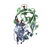
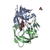
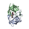
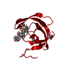
 Movie
Movie Controller

[English] 日本語
 Yorodumi
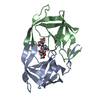
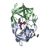
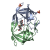
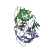
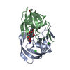
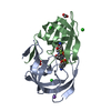
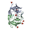
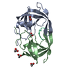
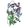
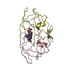
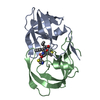
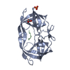
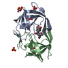
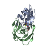
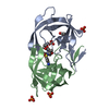
- PDB-6dh0: Crystal structure of HIV-1 Protease NL4-3 I84V Mutant in complex ... -
+ Open data

- Basic information
| Entry | Database: PDB / ID: 6dh0 | ||||||
|---|---|---|---|---|---|---|---|
| Title | Crystal structure of HIV-1 Protease NL4-3 I84V Mutant in complex with darunavir | ||||||
 Components Components | Protease | ||||||
 Keywords Keywords | HYDROLASE/HYDROLASE INHIBITOR / HIV / Protease / NL4-3 / DRUG RESISTANCE / PROTEASE INHIBITOR / HYDROLASE INHIBITOR COMPLEX / HYDROLASE / HYDROLASE-HYDROLASE INHIBITOR complex | ||||||
| Function / homology |  Function and homology information Function and homology information | ||||||
| Biological species |   Human immunodeficiency virus 1 Human immunodeficiency virus 1 | ||||||
| Method | X-RAY DIFFRACTION / MOLECULAR REPLACEMENT / Resolution: 1.899 Å | ||||||
 Authors Authors | Lockbaum, G.J. / Schiffer, C.A. | ||||||
| Funding support |  United States, 1items United States, 1items
| ||||||
 Citation Citation | Journal: ACS Infect Dis / Year: 2019 Title: Structural Adaptation of Darunavir Analogues against Primary Mutations in HIV-1 Protease. Authors: Lockbaum, G.J. / Leidner, F. / Rusere, L.N. / Henes, M. / Kosovrasti, K. / Nachum, G.S. / Nalivaika, E.A. / Ali, A. / Yilmaz, N.K. / Schiffer, C.A. | ||||||
| History |
|
- Structure visualization
| Structure viewer | Molecule: MolmilJmol/JSmol |
|---|
- Downloads & links
-Download
| PDBx/mmCIF format | 6dh0.cif.gz | 89.9 KB | Display | PDBx/mmCIF format |
|---|---|---|---|---|
| PDB format | pdb6dh0.ent.gz | 69 KB | Display | PDB format |
| PDBx/mmJSON format | 6dh0.json.gz | Tree view | PDBx/mmJSON format | |
| Others |  Other downloads Other downloads |
-Validation report
| Arichive directory | https://data.pdbj.org/pub/pdb/validation_reports/dh/6dh0ftp://data.pdbj.org/pub/pdb/validation_reports/dh/6dh0 | HTTPS FTP |
|---|
-Related structure data
| Related structure data |  6dgxC  6dgyC  6dgzC  6dh1C 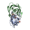 6dh2C 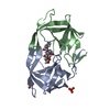 6dh3C  6dh4C 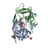 6dh5C 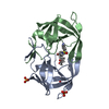 6dh6C  6dh7C  6dh8C  4ll3S S: Starting model for refinement C: citing same article ( |
|---|---|
| Similar structure data |

-Links
 PDBj
PDBj
- Assembly
| Deposited unit | 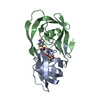
| ||||||||
|---|---|---|---|---|---|---|---|---|---|
| 1 |
| ||||||||
| Unit cell |
|
-Components
| #1: Protein | Mass: 10817.807 Da / Num. of mol.: 2 Source method: isolated from a genetically manipulated source Source: (gene. exp.) Human immunodeficiency virus 1 / Gene: pol / Plasmid: pXC35 / Production host:  Escherichia coli (E. coli) / References: UniProt: Q7ZCI0 Escherichia coli (E. coli) / References: UniProt: Q7ZCI0#2: Chemical | ChemComp-017 / ( | Darunavir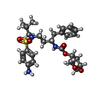  Mass: 547.664 Da / Num. of mol.: 1 / Source method: obtained synthetically / Formula: C27H37N3O7S / Feature type: SUBJECT OF INVESTIGATION / Comment: medication, antiretroviral*YM Mass: 547.664 Da / Num. of mol.: 1 / Source method: obtained synthetically / Formula: C27H37N3O7S / Feature type: SUBJECT OF INVESTIGATION / Comment: medication, antiretroviral*YM#3: Water | ChemComp-HOH / | Water Mass: 18.015 Da / Num. of mol.: 156 / Source method: isolated from a natural source / Formula: H2O Mass: 18.015 Da / Num. of mol.: 156 / Source method: isolated from a natural source / Formula: H2O |
|---|
-Experimental details
-Experiment
| Experiment | Method: X-RAY DIFFRACTION / Number of used crystals: 1 |
|---|
- Sample preparation
| Crystal | Density Matthews: 2.12 Å3/Da / Density % sol: 41.97 % |
|---|---|
| Crystal grow | Temperature: 293 K / Method: vapor diffusion, hanging drop Details: 23-24% (w/v) Ammonium Sulfate, 0.1M Bis-Tris-Methane-HCl Buffer pH 5.5 |
-Data collection
| Diffraction | Mean temperature: 100 K |
|---|---|
| Diffraction source | Source: ROTATING ANODE / Type: RIGAKU MICROMAX-007 HF / Wavelength: 1.54178 Å |
| Detector | Type: RIGAKU SATURN 944 / Detector: CCD / Date: Jun 20, 2016 |
| Radiation | Protocol: SINGLE WAVELENGTH / Monochromatic (M) / Laue (L): M / Scattering type: x-ray |
| Radiation wavelength | Wavelength: 1.54178 Å / Relative weight: 1 |
| Reflection | Resolution: 1.899→26.451 Å / Num. obs: 15103 / % possible obs: 99.9 % / Redundancy: 12 % / Net I/σ(I): 12.4 |
| Reflection shell | Resolution: 1.899→2.0451 Å |
- Processing
| Software |
| ||||||||||||||||||||||||||||||||||||||||||
|---|---|---|---|---|---|---|---|---|---|---|---|---|---|---|---|---|---|---|---|---|---|---|---|---|---|---|---|---|---|---|---|---|---|---|---|---|---|---|---|---|---|---|---|
| Refinement | Method to determine structure: MOLECULAR REPLACEMENT Starting model: 4LL3 Resolution: 1.899→26.451 Å / SU ML: 0.16 / Cross valid method: FREE R-VALUE / σ(F): 1.34 / Phase error: 22.93
| ||||||||||||||||||||||||||||||||||||||||||
| Solvent computation | Shrinkage radii: 0.9 Å / VDW probe radii: 1.11 Å | ||||||||||||||||||||||||||||||||||||||||||
| Refinement step | Cycle: LAST / Resolution: 1.899→26.451 Å
| ||||||||||||||||||||||||||||||||||||||||||
| Refine LS restraints |
| ||||||||||||||||||||||||||||||||||||||||||
| LS refinement shell |
|
