 Movie
Movie Controller

[English] 日本語
 Yorodumi
- PDB-1f7a: HOW DOES A SYMMETRIC DIMER RECOGNIZE AN ASYMMETRIC SUBSTRATE? A S... -
+ Open data

- Basic information
| Entry | Database: PDB / ID: 1f7a | ||||||
|---|---|---|---|---|---|---|---|
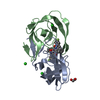
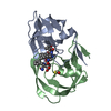
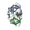
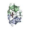
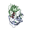
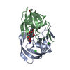
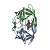
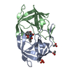
| Title | HOW DOES A SYMMETRIC DIMER RECOGNIZE AN ASYMMETRIC SUBSTRATE? A SUBSTRATE COMPLEX OF HIV-1 PROTEASE. | ||||||
 Components Components |
| ||||||
 Keywords Keywords |  HYDROLASE / CAPSID / SUBSTRATE RECOGNITION HYDROLASE / CAPSID / SUBSTRATE RECOGNITION | ||||||
| Function / homology |  Function and homology information Function and homology informationviral budding via host ESCRT complex / RNA stem-loop binding / host cell / HIV-1 retropepsin / retroviral ribonuclease H / exoribonuclease H / exoribonuclease H activity / host multivesicular body / DNA integration / RNA-directed DNA polymerase ...viral budding via host ESCRT complex / RNA stem-loop binding / host cell / HIV-1 retropepsin / retroviral ribonuclease H / exoribonuclease H / exoribonuclease H activity / host multivesicular body / DNA integration / RNA-directed DNA polymerase / viral genome integration into host DNA / viral penetration into host nucleus / establishment of integrated proviral latency / RNA-directed DNA polymerase activity / RNA-DNA hybrid ribonuclease activity / Transferases; Transferring phosphorus-containing groups; Nucleotidyltransferases / symbiont-mediated suppression of host gene expression / viral nucleocapsid / DNA recombination / Hydrolases; Acting on ester bonds / DNA-directed DNA polymerase / aspartic-type endopeptidase activity / DNA-directed DNA polymerase activity / symbiont entry into host cell / lipid binding / host cell nucleus / host cell plasma membrane / virion membrane / structural molecule activity / proteolysis / DNA binding / RNA binding / zinc ion binding / membrane / plasma membrane / cytoplasmSimilarity search - Function | ||||||
| Biological species |   Human immunodeficiency virus 1 Human immunodeficiency virus 1 | ||||||
| Method | X-RAY DIFFRACTION / MOLECULAR REPLACEMENT / Resolution: 2 Å | ||||||
 Authors Authors | Schiffer, C.A. | ||||||
 Citation Citation | Journal: J.Mol.Biol. / Year: 2000 Title: How does a symmetric dimer recognize an asymmetric substrate? A substrate complex of HIV-1 protease. Authors: Prabu-Jeyabalan, M. / Nalivaika, E. / Schiffer, C.A. | ||||||
| History |
|
- Structure visualization
| Structure viewer | Molecule: MolmilJmol/JSmol |
|---|
- Downloads & links
-Download
| PDBx/mmCIF format | 1f7a.cif.gz | 54.7 KB | Display | PDBx/mmCIF format |
|---|---|---|---|---|
| PDB format | pdb1f7a.ent.gz | 38.8 KB | Display | PDB format |
| PDBx/mmJSON format | 1f7a.json.gz | Tree view | PDBx/mmJSON format | |
| Others |  Other downloads Other downloads |
-Validation report
| Arichive directory | https://data.pdbj.org/pub/pdb/validation_reports/f7/1f7aftp://data.pdbj.org/pub/pdb/validation_reports/f7/1f7a | HTTPS FTP |
|---|
-Related structure data
| Related structure data |  1mtrS S: Starting model for refinement |
|---|---|
| Similar structure data |


-Links
 PDBj
PDBj


- Assembly
| Deposited unit | 
| ||||||||
|---|---|---|---|---|---|---|---|---|---|
| 1 |
| ||||||||
| Unit cell |
|
-Components
| #1: Protein | Mass: 10800.777 Da / Num. of mol.: 2 / Fragment: HIV-1 PROTEASE / Mutation: Q7K D25N Source method: isolated from a genetically manipulated source Source: (gene. exp.) Human immunodeficiency virus 1 / Genus: Lentivirus / Production host:  Escherichia coli (E. coli) / References: UniProt: P03369, HIV-1 retropepsin Escherichia coli (E. coli) / References: UniProt: P03369, HIV-1 retropepsin#2: Protein/peptide | | Mass: 1077.298 Da / Num. of mol.: 1 / Fragment: CA-P2 SUBSTRATE / Source method: obtained synthetically / Details: This peptide was chemically synthesized. / References: UniProt: Q9YX54*PLUS #3: Chemical | ChemComp-ACT / Acetate  Mass: 59.044 Da / Num. of mol.: 6 / Source method: obtained synthetically / Formula: C2H3O2 Mass: 59.044 Da / Num. of mol.: 6 / Source method: obtained synthetically / Formula: C2H3O2#4: Water | ChemComp-HOH / | Water Mass: 18.015 Da / Num. of mol.: 96 / Source method: isolated from a natural source / Formula: H2O Mass: 18.015 Da / Num. of mol.: 96 / Source method: isolated from a natural source / Formula: H2O |
|---|
-Experimental details
-Experiment
| Experiment | Method: X-RAY DIFFRACTION / Number of used crystals: 1 |
|---|
- Sample preparation
| Crystal | Density Matthews: 2.06 Å3/Da / Density % sol: 40.28 % | ||||||||||||||||||||||||||||||
|---|---|---|---|---|---|---|---|---|---|---|---|---|---|---|---|---|---|---|---|---|---|---|---|---|---|---|---|---|---|---|---|
| Crystal grow | *PLUS pH: 6.2 / Method: vapor diffusion, hanging drop | ||||||||||||||||||||||||||||||
| Components of the solutions | *PLUS
|
-Data collection
| Diffraction | Mean temperature: 298 K |
|---|---|
| Diffraction source | Source: ROTATING ANODE / Type: RIGAKU / Wavelength: 1.5418 / Wavelength: 1.5418 Å |
| Detector | Type: RIGAKU RAXIS IV / Detector: IMAGE PLATE / Date: Dec 1, 1998 |
| Radiation | Protocol: SINGLE WAVELENGTH / Monochromatic (M) / Laue (L): M / Scattering type: x-ray |
| Radiation wavelength | Wavelength: 1.5418 Å / Relative weight: 1 |
| Reflection | Resolution: 2→50 Å / Num. all: 49525 / Num. obs: 12474 / % possible obs: 94.3 % / Redundancy: 5 % / Biso Wilson estimate: 20.6 Å2 / Rmerge(I) obs: 0.048 / Net I/σ(I): 15.4 |
| Reflection shell | Resolution: 2→2.05 Å / Redundancy: 5 % / Rmerge(I) obs: 0.213 / Num. unique all: 738 / Rsym value: 21.3 / % possible all: 84 |
| Reflection | *PLUS Num. measured all: 49525 |
- Processing
| Software |
| |||||||||||||||||||||||||
|---|---|---|---|---|---|---|---|---|---|---|---|---|---|---|---|---|---|---|---|---|---|---|---|---|---|---|
| Refinement | Method to determine structure: MOLECULAR REPLACEMENT Starting model: 1MTR Resolution: 2→26.37 Å / Rfactor Rfree error: 0.007 / Data cutoff high absF: 304680.17 / Data cutoff low absF: 0 / Isotropic thermal model: RESTRAINED / Cross valid method: THROUGHOUT / σ(F): 0 / σ(I): 0
| |||||||||||||||||||||||||
| Solvent computation | Solvent model: FLAT MODEL / Bsol: 74.89 Å2 / ksol: 0.349 e/Å3 | |||||||||||||||||||||||||
| Displacement parameters | Biso mean: 32.9 Å2
| |||||||||||||||||||||||||
| Refine analyze |
| |||||||||||||||||||||||||
| Refinement step | Cycle: LAST / Resolution: 2→26.37 Å
| |||||||||||||||||||||||||
| Refine LS restraints |
| |||||||||||||||||||||||||
| LS refinement shell | Resolution: 2→2.1 Å / Rfactor Rfree error: 0.024 / Total num. of bins used: 6
| |||||||||||||||||||||||||
| Xplor file |
| |||||||||||||||||||||||||
| Software | *PLUS Name: CNS / Version: 0.9 / Classification: refinement | |||||||||||||||||||||||||
| Refinement | *PLUS σ(F): 0 / % reflection Rfree: 10.4 % / Rfactor obs: 0.197 | |||||||||||||||||||||||||
| Solvent computation | *PLUS | |||||||||||||||||||||||||
| Displacement parameters | *PLUS Biso mean: 32.9 Å2 | |||||||||||||||||||||||||
| Refine LS restraints | *PLUS
| |||||||||||||||||||||||||
| LS refinement shell | *PLUS Highest resolution: 2 Å / Lowest resolution: 2.1 Å / % reflection Rfree: 10.4 % |
