 Movie
Movie Controller


+ Open data

- Basic information
| Entry | Database: PDB / ID: 3d1z | ||||||
|---|---|---|---|---|---|---|---|
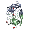
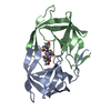
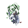
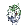
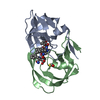
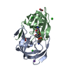
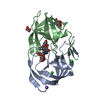
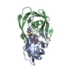
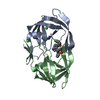
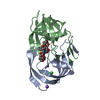
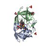
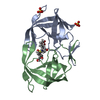
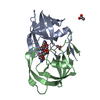
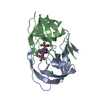
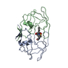
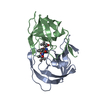
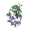
| Title | Crystal structure of HIV-1 mutant I54M and inhibitor DARUNAVIR | ||||||
 Components Components | HIV-1 Protease | ||||||
 Keywords Keywords | HYDROLASE / DRUG RESISTANCE / HIV-1 / flap mutant / I54M / Darunavir | ||||||
| Function / homology |  Function and homology information Function and homology informationRNA stem-loop binding / host cell / HIV-1 retropepsin / retroviral ribonuclease H / exoribonuclease H / exoribonuclease H activity / host multivesicular body / DNA integration / RNA-directed DNA polymerase / viral genome integration into host DNA ...RNA stem-loop binding / host cell / HIV-1 retropepsin / retroviral ribonuclease H / exoribonuclease H / exoribonuclease H activity / host multivesicular body / DNA integration / RNA-directed DNA polymerase / viral genome integration into host DNA / viral penetration into host nucleus / establishment of integrated proviral latency / RNA-directed DNA polymerase activity / RNA-DNA hybrid ribonuclease activity / Transferases; Transferring phosphorus-containing groups; Nucleotidyltransferases / symbiont-mediated suppression of host gene expression / viral nucleocapsid / DNA recombination / Hydrolases; Acting on ester bonds / DNA-directed DNA polymerase / aspartic-type endopeptidase activity / DNA-directed DNA polymerase activity / symbiont entry into host cell / lipid binding / host cell nucleus / host cell plasma membrane / virion membrane / structural molecule activity / proteolysis / DNA binding / zinc ion binding / membraneSimilarity search - Function | ||||||
| Biological species |   Human immunodeficiency virus type 1 Human immunodeficiency virus type 1 | ||||||
| Method | X-RAY DIFFRACTION / SYNCHROTRON / MOLECULAR REPLACEMENT / Resolution: 1.3 Å | ||||||
 Authors Authors | Liu, F. / Kovalesky, A.Y. / Tie, Y. / Ghosh, A.K. / Harrison, R.W. / Weber, I.T. | ||||||
 Citation Citation | Journal: J.Mol.Biol. / Year: 2008 Title: Effect of flap mutations on structure of HIV-1 protease and inhibition by saquinavir and darunavir. Authors: Liu, F. / Kovalevsky, A.Y. / Tie, Y. / Ghosh, A.K. / Harrison, R.W. / Weber, I.T. #1: Journal: J.Mol.Biol. / Year: 2006Title: Ultra-High Resolution Crystal Structure of HIV-1 Protease Mutant Reveals Two Binding Sites for Clinical Inhibitor Tmc114 Authors: Kovalevsky, A.Y. / Liu, F. / Leshchenko, S. / Ghosh, A.K. / Harrison, R.W. / Weber, I.T. #2: Journal: J.Mol.Biol. / Year: 2005Title: Kinetic, Stability, and Structural Changes in High-Resolution Crystal Structures of HIV-1 Protease with Drug-Resistant Mutations L24I, I50V, and G73S. Authors: Liu, F. / Boross, P.I. / Wang, Y.F. / Tozser, J. / Louis, J.M. / Harrison, R.W. / Weber, I.T. | ||||||
| History |
|
- Structure visualization
| Structure viewer | Molecule: MolmilJmol/JSmol |
|---|
- Downloads & links
-Download
| PDBx/mmCIF format | 3d1z.cif.gz | 107.4 KB | Display | PDBx/mmCIF format |
|---|---|---|---|---|
| PDB format | pdb3d1z.ent.gz | 82.4 KB | Display | PDB format |
| PDBx/mmJSON format | 3d1z.json.gz | Tree view | PDBx/mmJSON format | |
| Others |  Other downloads Other downloads |
-Validation report
| Arichive directory | https://data.pdbj.org/pub/pdb/validation_reports/d1/3d1zftp://data.pdbj.org/pub/pdb/validation_reports/d1/3d1z | HTTPS FTP |
|---|
-Related structure data
| Related structure data |  3cywC  3cyxC  3d1xC 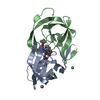 3d1yC  3d20C 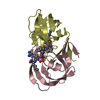 1dazS C: citing same article ( S: Starting model for refinement |
|---|---|
| Similar structure data |


-Links
 PDBj
PDBj


- Assembly
| Deposited unit | 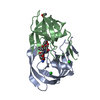
| ||||||||
|---|---|---|---|---|---|---|---|---|---|
| 1 |
| ||||||||
| Unit cell |
|
-Components
| #1: Protein | / Retropepsin / PR Mass: 10758.715 Da / Num. of mol.: 2 / Mutation: I54M Source method: isolated from a genetically manipulated source Source: (gene. exp.) Human immunodeficiency virus type 1 / Gene: gag-pol / Production host:  ESCHERICHIA COLI (E. coli) / References: UniProt: P04587, HIV-1 retropepsin ESCHERICHIA COLI (E. coli) / References: UniProt: P04587, HIV-1 retropepsin#2: Chemical | Chloride  Mass: 35.453 Da / Num. of mol.: 3 / Source method: obtained synthetically / Formula: Cl Mass: 35.453 Da / Num. of mol.: 3 / Source method: obtained synthetically / Formula: Cl#3: Chemical | ChemComp-017 / ( | Darunavir  Mass: 547.664 Da / Num. of mol.: 1 / Source method: obtained synthetically / Formula: C27H37N3O7S / Comment: medication, antiretroviral*YM Mass: 547.664 Da / Num. of mol.: 1 / Source method: obtained synthetically / Formula: C27H37N3O7S / Comment: medication, antiretroviral*YM#4: Chemical | ChemComp-ACY / | Acetic acid  Mass: 60.052 Da / Num. of mol.: 1 / Source method: obtained synthetically / Formula: C2H4O2 Mass: 60.052 Da / Num. of mol.: 1 / Source method: obtained synthetically / Formula: C2H4O2#5: Water | ChemComp-HOH / | Water Mass: 18.015 Da / Num. of mol.: 188 / Source method: isolated from a natural source / Formula: H2O Mass: 18.015 Da / Num. of mol.: 188 / Source method: isolated from a natural source / Formula: H2OSequence details | MUTATIONS Q7K, L33I, L63I, C67A, C95A, HAVE BEEN MADE TO STABILIZE THE PROTEASE FROM ...MUTATIONS Q7K, L33I, L63I, C67A, C95A, HAVE BEEN MADE TO STABILIZE THE PROTEASE FROM AUTOPROTEO | |
|---|
-Experimental details
-Experiment
| Experiment | Method: X-RAY DIFFRACTION / Number of used crystals: 1 |
|---|
- Sample preparation
| Crystal | Density Matthews: 2.68 Å3/Da / Density % sol: 54.07 % |
|---|---|
| Crystal grow | Temperature: 295 K / Method: vapor diffusion, hanging drop / pH: 4.6 Details: SODIUM ACETATE BUFFER, 20-25% NaCl, pH 4.6, VAPOR DIFFUSION, HANGING DROP, temperature 295K |
-Data collection
| Diffraction | Mean temperature: 95 K |
|---|---|
| Diffraction source | Source: SYNCHROTRON / Site: APS  / Beamline: 22-ID / Wavelength: 1 / Beamline: 22-ID / Wavelength: 1 |
| Detector | Type: MARMOSAIC 300 mm CCD / Detector: CCD / Date: Jun 12, 2005 |
| Radiation | Monochromator: SI / Protocol: SINGLE WAVELENGTH / Monochromatic (M) / Laue (L): M / Scattering type: x-ray |
| Radiation wavelength | Wavelength: 1 Å / Relative weight: 1 |
| Reflection | Resolution: 1.3→50 Å / Num. all: 53524 / Num. obs: 43720 / % possible obs: 81.7 % / Observed criterion σ(F): 4 / Observed criterion σ(I): 2 / Redundancy: 5.7 % / Rmerge(I) obs: 0.089 / Net I/σ(I): 24.4 |
| Reflection shell | Resolution: 1.3→1.35 Å / Redundancy: 2.6 % / Rmerge(I) obs: 0.57 / Mean I/σ(I) obs: 2.2 / % possible all: 80.8 |
- Processing
| Software |
| |||||||||||||||||||||||||||||||||
|---|---|---|---|---|---|---|---|---|---|---|---|---|---|---|---|---|---|---|---|---|---|---|---|---|---|---|---|---|---|---|---|---|---|---|
| Refinement | Method to determine structure: MOLECULAR REPLACEMENT Starting model: PDB entry 1DAZ Resolution: 1.3→10 Å / Num. parameters: 17146 / Num. restraintsaints: 22351 / Cross valid method: FREE R / σ(F): 0 / Stereochemistry target values: ENGH AND HUBER / Details: ANISOTROPIC REFINEMENT REDUCED FREE R (NO CUTOFF)
| |||||||||||||||||||||||||||||||||
| Refine analyze | Num. disordered residues: 29 / Occupancy sum hydrogen: 1626 / Occupancy sum non hydrogen: 1715.5 | |||||||||||||||||||||||||||||||||
| Refinement step | Cycle: LAST / Resolution: 1.3→10 Å
| |||||||||||||||||||||||||||||||||
| Refine LS restraints |
|
