 Movie
Movie Controller

[English] 日本語
 Yorodumi
- PDB-6c5r: Crystal structure of the soluble domain of the mitochondrial calc... -
+ Open data

- Basic information
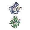
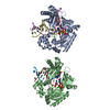
| Entry | Database: PDB / ID: 6c5r | ||||||
|---|---|---|---|---|---|---|---|
| Title | Crystal structure of the soluble domain of the mitochondrial calcium uniporter | ||||||
 Components Components | calcium uniporter | ||||||
 Keywords Keywords |  CYTOSOLIC PROTEIN CYTOSOLIC PROTEIN | ||||||
| Function / homology | uniporter activity / Calcium uniporter protein, C-terminal / MCU family / Mitochondrial calcium uniporter / mitochondrial calcium ion homeostasis / calcium channel activity / mitochondrial inner membrane / identical protein binding / Calcium uniporter protein Function and homology information Function and homology information | ||||||
| Biological species |  Metarhizium acridum (fungus) Metarhizium acridum (fungus) | ||||||
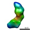
| Method | X-RAY DIFFRACTION / SYNCHROTRON / Resolution: 3.09608311048 Å | ||||||
 Authors Authors | Fan, C. / Fan, M. / Fastman, N. / Zhang, J. / Feng, L. | ||||||
 Citation Citation | Journal: Nature / Year: 2018 Title: X-ray and cryo-EM structures of the mitochondrial calcium uniporter. Authors: Chao Fan / Minrui Fan / Benjamin J Orlando / Nathan M Fastman / Jinru Zhang / Yan Xu / Melissa G Chambers / Xiaofang Xu / Kay Perry / Maofu Liao / Liang Feng /   Abstract: Mitochondrial calcium uptake is critical for regulating ATP production, intracellular calcium signalling, and cell death. This uptake is mediated by a highly selective calcium channel called the ...Mitochondrial calcium uptake is critical for regulating ATP production, intracellular calcium signalling, and cell death. This uptake is mediated by a highly selective calcium channel called the mitochondrial calcium uniporter (MCU). Here, we determined the structures of the pore-forming MCU proteins from two fungi by X-ray crystallography and single-particle cryo-electron microscopy. The stoichiometry, overall architecture, and individual subunit structure differed markedly from those described in the recent nuclear magnetic resonance structure of Caenorhabditis elegans MCU. We observed a dimer-of-dimer architecture across species and chemical environments, which was corroborated by biochemical experiments. Structural analyses and functional characterization uncovered the roles of key residues in the pore. These results reveal a new ion channel architecture, provide insights into calcium coordination, selectivity and conduction, and establish a structural framework for understanding the mechanism of mitochondrial calcium uniporter function. | ||||||
| History |
|
- Structure visualization
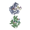
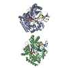
| Structure viewer | Molecule: MolmilJmol/JSmol |
|---|
- Downloads & links
-Download
| PDBx/mmCIF format | 6c5r.cif.gz | 316.2 KB | Display | PDBx/mmCIF format |
|---|---|---|---|---|
| PDB format | pdb6c5r.ent.gz | 208.3 KB | Display | PDB format |
| PDBx/mmJSON format | 6c5r.json.gz | Tree view | PDBx/mmJSON format | |
| Others |  Other downloads Other downloads |
-Validation report
| Arichive directory | https://data.pdbj.org/pub/pdb/validation_reports/c5/6c5rftp://data.pdbj.org/pub/pdb/validation_reports/c5/6c5r | HTTPS FTP |
|---|
-Related structure data
| Related structure data | 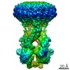 7800C 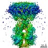 7801C 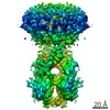 7802C 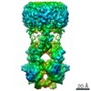 7803C 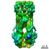 7804C  6c5wC C: citing same article ( |
|---|---|
| Similar structure data |


-Links
 PDBj
PDBj- Assembly
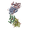
| Deposited unit | 
| ||||||||||||
|---|---|---|---|---|---|---|---|---|---|---|---|---|---|
| 1 | 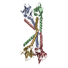
| ||||||||||||
| 2 | 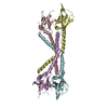
| ||||||||||||
| Unit cell |
|
-Components
| #1: Protein | Mass: 24267.676 Da / Num. of mol.: 8 Source method: isolated from a genetically manipulated source Source: (gene. exp.) Metarhizium acridum (strain CQMa 102) (fungus)Strain: CQMa 102 / Gene: MAC_01752 / Production host:  Escherichia coli K-12 (bacteria) / References: UniProt: E9DVV4 Escherichia coli K-12 (bacteria) / References: UniProt: E9DVV4 |
|---|
-Experimental details
-Experiment
| Experiment | Method: X-RAY DIFFRACTION / Number of used crystals: 1 |
|---|
- Sample preparation
| Crystal | Density Matthews: 3.43 Å3/Da / Density % sol: 64.1 % |
|---|---|
| Crystal grow | Temperature: 293 K / Method: vapor diffusion, hanging drop Details: 0.1 M glycine, pH 9.3, 15.5% PEG 4000, and 0.6 M NaNO3 |
-Data collection
| Diffraction | Mean temperature: 100 K |
|---|---|
| Diffraction source | Source: SYNCHROTRON / Site: APS / Beamline: 17-ID / Wavelength: 1 Å |
| Detector | Type: DECTRIS PILATUS3 S 6M / Detector: PIXEL / Date: Mar 25, 2017 |
| Radiation | Protocol: SINGLE WAVELENGTH / Monochromatic (M) / Laue (L): M / Scattering type: x-ray |
| Radiation wavelength | Wavelength: 1 Å / Relative weight: 1 |
| Reflection | Resolution: 3.09608311048→50 Å / Num. obs: 47337 / % possible obs: 100 % / Redundancy: 3.4 % / Biso Wilson estimate: 97.3071351599 Å2 / Rmerge(I) obs: 0.089 / Net I/σ(I): 14.1 |
| Reflection shell | Resolution: 3.1→3.17 Å / Num. unique obs: 3153 |
- Processing
| Software |
| |||||||||||||||||||||||||||||||||||||||||||||||||||||||||||||||||||||||||||||||||||||||||||||||||||||||||
|---|---|---|---|---|---|---|---|---|---|---|---|---|---|---|---|---|---|---|---|---|---|---|---|---|---|---|---|---|---|---|---|---|---|---|---|---|---|---|---|---|---|---|---|---|---|---|---|---|---|---|---|---|---|---|---|---|---|---|---|---|---|---|---|---|---|---|---|---|---|---|---|---|---|---|---|---|---|---|---|---|---|---|---|---|---|---|---|---|---|---|---|---|---|---|---|---|---|---|---|---|---|---|---|---|---|---|
| Refinement | Resolution: 3.09608311048→42.105090976 Å / SU ML: 0.448428869909 / Cross valid method: FREE R-VALUE / σ(F): 1.33787117744 / Phase error: 34.267652959 Stereochemistry target values: GeoStd + Monomer Library + CDL v1.2
| |||||||||||||||||||||||||||||||||||||||||||||||||||||||||||||||||||||||||||||||||||||||||||||||||||||||||
| Solvent computation | Shrinkage radii: 0.9 Å / VDW probe radii: 1.11 Å / Solvent model: FLAT BULK SOLVENT MODEL | |||||||||||||||||||||||||||||||||||||||||||||||||||||||||||||||||||||||||||||||||||||||||||||||||||||||||
| Displacement parameters | Biso mean: 99.0706387757 Å2 | |||||||||||||||||||||||||||||||||||||||||||||||||||||||||||||||||||||||||||||||||||||||||||||||||||||||||
| Refinement step | Cycle: LAST / Resolution: 3.09608311048→42.105090976 Å
| |||||||||||||||||||||||||||||||||||||||||||||||||||||||||||||||||||||||||||||||||||||||||||||||||||||||||
| Refine LS restraints |
| |||||||||||||||||||||||||||||||||||||||||||||||||||||||||||||||||||||||||||||||||||||||||||||||||||||||||
| LS refinement shell |
|
