 Movie
Movie Controller

[English] 日本語
 Yorodumi
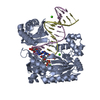
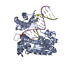
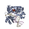
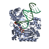
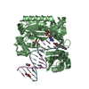
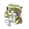
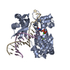
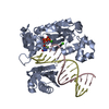
- PDB-3gij: Dpo4 extension ternary complex with oxoG(syn)-A(anti) and oxoG(an... -
+ Open data

- Basic information
| Entry | Database: PDB / ID: 3gij | ||||||
|---|---|---|---|---|---|---|---|
| Title | Dpo4 extension ternary complex with oxoG(syn)-A(anti) and oxoG(anti)-A(syn) pairs | ||||||
 Components Components |
| ||||||
 Keywords Keywords | TRANSFERASE/DNA /  DNA POLYMERASE / 8-OXOGUANINE / Y-FAMILY / LESION BYPASS / DNA damage / DNA repair / DNA replication / DNA-binding / DNA-directed DNA polymerase / Magnesium / Metal-binding / Mutator protein / Nucleotidyltransferase / Transferase / TRANSFERASE-DNA COMPLEX DNA POLYMERASE / 8-OXOGUANINE / Y-FAMILY / LESION BYPASS / DNA damage / DNA repair / DNA replication / DNA-binding / DNA-directed DNA polymerase / Magnesium / Metal-binding / Mutator protein / Nucleotidyltransferase / Transferase / TRANSFERASE-DNA COMPLEX | ||||||
| Function / homology |  Function and homology information Function and homology informationerror-prone translesion synthesis / DNA-templated DNA replication / damaged DNA binding / DNA-directed DNA polymerase / DNA-directed DNA polymerase activity / magnesium ion binding / cytoplasmSimilarity search - Function | ||||||
| Biological species |   Sulfolobus solfataricus P2 (archaea) Sulfolobus solfataricus P2 (archaea) | ||||||
| Method | X-RAY DIFFRACTION / SYNCHROTRON / MOLECULAR REPLACEMENT / Resolution: 2.4 Å | ||||||
 Authors Authors | Rechkoblit, O. / Malinina, L. / Patel, D.J. | ||||||
 Citation Citation | Journal: Structure / Year: 2009 Title: Impact of conformational heterogeneity of OxoG lesions and their pairing partners on bypass fidelity by Y family polymerases. Authors: Rechkoblit, O. / Malinina, L. / Cheng, Y. / Geacintov, N.E. / Broyde, S. / Patel, D.J. | ||||||
| History |
|
- Structure visualization
| Structure viewer | Molecule: MolmilJmol/JSmol |
|---|
- Downloads & links
-Download
| PDBx/mmCIF format | 3gij.cif.gz | 193.9 KB | Display | PDBx/mmCIF format |
|---|---|---|---|---|
| PDB format | pdb3gij.ent.gz | 146.7 KB | Display | PDB format |
| PDBx/mmJSON format | 3gij.json.gz | Tree view | PDBx/mmJSON format | |
| Others |  Other downloads Other downloads |
-Validation report
| Arichive directory | https://data.pdbj.org/pub/pdb/validation_reports/gi/3gijftp://data.pdbj.org/pub/pdb/validation_reports/gi/3gij | HTTPS FTP |
|---|
-Related structure data
| Related structure data | 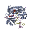 3giiSC  3gikC 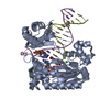 3gilC 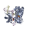 3gimC S: Starting model for refinement C: citing same article ( |
|---|---|
| Similar structure data |


-Links
 PDBj
PDBj


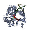
- Assembly
| Deposited unit | 
| ||||||||
|---|---|---|---|---|---|---|---|---|---|
| 1 | 
| ||||||||
| 2 | 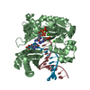
| ||||||||
| Unit cell |
|
-Components
-Protein , 1 types, 2 molecules AB
| #1: Protein | / Pol IV Mass: 38945.379 Da / Num. of mol.: 2 Source method: isolated from a genetically manipulated source Source: (gene. exp.) Sulfolobus solfataricus P2 (archaea) / Strain: P2 / DSM 1617 / JCM 11322 / Gene: dbh, dpo4, SSO2448 / Plasmid: pET28a / Production host:  Escherichia coli (E. coli) / Strain (production host): BL21(DE3)-RIL(STRATAGENE) / References: UniProt: Q97W02, DNA-directed DNA polymerase Escherichia coli (E. coli) / Strain (production host): BL21(DE3)-RIL(STRATAGENE) / References: UniProt: Q97W02, DNA-directed DNA polymerase |
|---|
-DNA chain , 2 types, 4 molecules DHEJ
| #2: DNA chain | Mass: 4070.668 Da / Num. of mol.: 2 / Source method: obtained synthetically / Details: DNA primer strand #3: DNA chain | Mass: 5405.521 Da / Num. of mol.: 2 / Source method: obtained synthetically / Details: DNA OXOG-modified template strand |
|---|
-Non-polymers , 3 types, 252 molecules 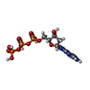


| #4: Chemical | Deoxyguanosine triphosphate Mass: 507.181 Da / Num. of mol.: 2 / Source method: obtained synthetically / Formula: C10H16N5O13P3 Mass: 507.181 Da / Num. of mol.: 2 / Source method: obtained synthetically / Formula: C10H16N5O13P3#5: Chemical | ChemComp-CA /  Mass: 40.078 Da / Num. of mol.: 6 / Source method: obtained synthetically / Formula: Ca Mass: 40.078 Da / Num. of mol.: 6 / Source method: obtained synthetically / Formula: Ca#6: Water | ChemComp-HOH / | WaterMass: 18.015 Da / Num. of mol.: 244 / Source method: isolated from a natural source / Formula: H2O |
|---|
-Experimental details
-Experiment
| Experiment | Method: X-RAY DIFFRACTION / Number of used crystals: 1 |
|---|
- Sample preparation
| Crystal | Density Matthews: 2.74 Å3/Da / Density % sol: 55.03 % | ||||||||||||||||
|---|---|---|---|---|---|---|---|---|---|---|---|---|---|---|---|---|---|
| Crystal grow | Temperature: 293 K / Method: vapor diffusion, hanging drop / pH: 7 Details: 100 mM HEPES, 100 mM Calcium acetate, 10% PEG 4000, pH 7.0, VAPOR DIFFUSION, HANGING DROP, temperature 293K | ||||||||||||||||
| Components of the solutions |
|
-Data collection
| Diffraction | Mean temperature: 100 K |
|---|---|
| Diffraction source | Source: SYNCHROTRON / Site: APS  / Beamline: 24-ID-C / Wavelength: 0.97942 Å / Beamline: 24-ID-C / Wavelength: 0.97942 Å |
| Detector | Type: ADSC QUANTUM 315 / Detector: CCD / Date: Nov 22, 2005 Details: 6144 x 6144 pixel elements with an effective pixel size less than 60x60 micron and resolution of order 90x90 micron |
| Radiation | Protocol: SINGLE WAVELENGTH / Monochromatic (M) / Laue (L): M / Scattering type: x-ray |
| Radiation wavelength | Wavelength: 0.97942 Å / Relative weight: 1 |
| Reflection | Resolution: 2.35→20 Å / Num. obs: 40120 / % possible obs: 92.7 % / Redundancy: 3.8 % / Rmerge(I) obs: 0.064 / Net I/σ(I): 20.1 |
| Reflection shell | Resolution: 2.35→2.42 Å / Rmerge(I) obs: 0.424 / Mean I/σ(I) obs: 1.8 / % possible all: 62.4 |
- Processing
| Software |
| |||||||||||||||||||||||||||||||||||||||||||||||||||||||||||||||||||||||||||||||||||||||||||||||||||||||||||||||||||||||||||||
|---|---|---|---|---|---|---|---|---|---|---|---|---|---|---|---|---|---|---|---|---|---|---|---|---|---|---|---|---|---|---|---|---|---|---|---|---|---|---|---|---|---|---|---|---|---|---|---|---|---|---|---|---|---|---|---|---|---|---|---|---|---|---|---|---|---|---|---|---|---|---|---|---|---|---|---|---|---|---|---|---|---|---|---|---|---|---|---|---|---|---|---|---|---|---|---|---|---|---|---|---|---|---|---|---|---|---|---|---|---|---|---|---|---|---|---|---|---|---|---|---|---|---|---|---|---|---|
| Refinement | Method to determine structure: MOLECULAR REPLACEMENT Starting model: PDB entry 3GII Resolution: 2.4→20 Å / Cor.coef. Fo:Fc: 0.944 / Cor.coef. Fo:Fc free: 0.925 / Occupancy max: 1 / Occupancy min: 0.2 / SU B: 17.44 / SU ML: 0.215 / TLS residual ADP flag: LIKELY RESIDUAL / Cross valid method: THROUGHOUT / σ(F): 0 / ESU R: 0.472 / ESU R Free: 0.276 / Stereochemistry target values: MAXIMUM LIKELIHOOD / Details: HYDROGENS HAVE BEEN ADDED IN THE RIDING POSITIONS
| |||||||||||||||||||||||||||||||||||||||||||||||||||||||||||||||||||||||||||||||||||||||||||||||||||||||||||||||||||||||||||||
| Solvent computation | Ion probe radii: 0.8 Å / Shrinkage radii: 0.8 Å / VDW probe radii: 1.2 Å / Solvent model: MASK | |||||||||||||||||||||||||||||||||||||||||||||||||||||||||||||||||||||||||||||||||||||||||||||||||||||||||||||||||||||||||||||
| Displacement parameters | Biso max: 102.64 Å2 / Biso mean: 61.17 Å2 / Biso min: 17.18 Å2
| |||||||||||||||||||||||||||||||||||||||||||||||||||||||||||||||||||||||||||||||||||||||||||||||||||||||||||||||||||||||||||||
| Refinement step | Cycle: LAST / Resolution: 2.4→20 Å
| |||||||||||||||||||||||||||||||||||||||||||||||||||||||||||||||||||||||||||||||||||||||||||||||||||||||||||||||||||||||||||||
| Refine LS restraints |
| |||||||||||||||||||||||||||||||||||||||||||||||||||||||||||||||||||||||||||||||||||||||||||||||||||||||||||||||||||||||||||||
| LS refinement shell | Resolution: 2.4→2.461 Å / Total num. of bins used: 20
| |||||||||||||||||||||||||||||||||||||||||||||||||||||||||||||||||||||||||||||||||||||||||||||||||||||||||||||||||||||||||||||
| Refinement TLS params. | Method: refined / Refine-ID: X-RAY DIFFRACTION
| |||||||||||||||||||||||||||||||||||||||||||||||||||||||||||||||||||||||||||||||||||||||||||||||||||||||||||||||||||||||||||||
| Refinement TLS group |
|
