SOS response / error-prone translesion synthesis / DNA-templated DNA replication / damaged DNA binding / DNA-directed DNA polymerase / DNA-directed DNA polymerase activity / magnesium ion binding / cytosol Similarity search - Function
DNA polymerase type-Y, HhH motif / IMS family HHH motif / DNA polymerase IV / DNA polymerase, Y-family, little finger domain / MutS, DNA mismatch repair protein, domain I - #60 / MutS, DNA mismatch repair protein, domain I / DNA polymerase, Y-family, little finger domain / impB/mucB/samB family C-terminal domain / UmuC domain / DNA polymerase, Y-family, little finger domain superfamily ...DNA polymerase type-Y, HhH motif / IMS family HHH motif / DNA polymerase IV / DNA polymerase, Y-family, little finger domain / MutS, DNA mismatch repair protein, domain I - #60 / MutS, DNA mismatch repair protein, domain I / DNA polymerase, Y-family, little finger domain / impB/mucB/samB family C-terminal domain / UmuC domain / DNA polymerase, Y-family, little finger domain superfamily / impB/mucB/samB family / UmuC domain profile. / Reverse transcriptase/Diguanylate cyclase domain / Dna Ligase; domain 1 / 5' to 3' exonuclease, C-terminal subdomain / DNA polymerase; domain 1 / Reverse transcriptase/Diguanylate cyclase domain / Alpha-Beta Plaits / DNA/RNA polymerase superfamily / 2-Layer Sandwich / Orthogonal Bundle / 3-Layer(aba) Sandwich / Mainly Alpha / Alpha Beta Similarity search - Domain/homology
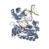
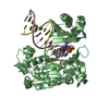
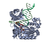
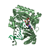
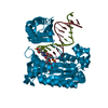
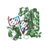
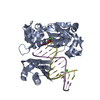
D: 5'-D(*GP*GP*TP*TP*GP*GP*AP*TP*GP*GP*TP*AP*(DDG))-3' E: 5'-D(*CP*TP*AP*AP*CP*(8OG)*CP*TP*AP*CP*CP*AP*TP*CP*CP*AP*AP*CP*C)-3' H: 5'-D(*GP*GP*TP*TP*GP*GP*AP*TP*GP*GP*TP*AP*(DDG))-3' J: 5'-D(*CP*TP*AP*AP*CP*(8OG)*CP*TP*AP*CP*CP*AP*TP*CP*CP*AP*AP*CP*C)-3' A: DNA polymerase IV B: DNA polymerase IV hetero molecules
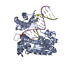
D: 5'-D(*GP*GP*TP*TP*GP*GP*AP*TP*GP*GP*TP*AP*(DDG))-3' E: 5'-D(*CP*TP*AP*AP*CP*(8OG)*CP*TP*AP*CP*CP*AP*TP*CP*CP*AP*AP*CP*C)-3' A: DNA polymerase IV hetero molecules
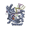
H: 5'-D(*GP*GP*TP*TP*GP*GP*AP*TP*GP*GP*TP*AP*(DDG))-3' J: 5'-D(*CP*TP*AP*AP*CP*(8OG)*CP*TP*AP*CP*CP*AP*TP*CP*CP*AP*AP*CP*C)-3' B: DNA polymerase IV hetero molecules
Resolution: 2.35→20 Å / Cor.coef. Fo:Fc: 0.93 / Cor.coef. Fo:Fc free: 0.881 / SU B: 30.371 / SU ML: 0.347 / TLS residual ADP flag: LIKELY RESIDUAL / Cross valid method: THROUGHOUT / ESU R: 0.653 / ESU R Free: 0.358 / Stereochemistry target values: MAXIMUM LIKELIHOOD Details: HYDROGENS HAVE BEEN ADDED IN THE RIDING POSITIONS. The detwinned data is used for refinement. The twinning fraction is 0.4, detwinning operator is l, -k, h.
Rfactor
Num. reflection
% reflection
Selection details
Rfree
0.31751
1750
5.1 %
RANDOM
Rwork
0.24557
-
-
-
obs
0.24909
32562
88.74 %
-
all
-
36438
-
-
Solvent computation
Ion probe radii: 0.8 Å / Shrinkage radii: 0.8 Å / VDW probe radii: 1.2 Å / Solvent model: MASK
Displacement parameters
Biso mean: 54.44 Å2
Baniso -1
Baniso -2
Baniso -3
1-
3.48 Å2
0 Å2
-1.21 Å2
2-
-
-1.97 Å2
0 Å2
3-
-
-
-2.24 Å2
Refinement step
Cycle: LAST / Resolution: 2.35→20 Å
Protein
Nucleic acid
Ligand
Solvent
Total
Num. atoms
5478
1052
3
144
6677
Refine LS restraints
Refine-ID
Type
Dev ideal
Dev ideal target
Number
X-RAY DIFFRACTION
r_bond_refined_d
0.01
0.022
6737
X-RAY DIFFRACTION
r_bond_other_d
X-RAY DIFFRACTION
r_angle_refined_deg
1.381
2.189
9276
X-RAY DIFFRACTION
r_angle_other_deg
X-RAY DIFFRACTION
r_dihedral_angle_1_deg
5.97
5
680
X-RAY DIFFRACTION
r_dihedral_angle_2_deg
37.876
23.636
242
X-RAY DIFFRACTION
r_dihedral_angle_3_deg
19.951
15
1120
X-RAY DIFFRACTION
r_dihedral_angle_4_deg
16.293
15
48
X-RAY DIFFRACTION
r_chiral_restr
0.081
0.2
1056
X-RAY DIFFRACTION
r_gen_planes_refined
0.003
0.02
4566
X-RAY DIFFRACTION
r_gen_planes_other
X-RAY DIFFRACTION
r_nbd_refined
0.212
0.2
2770
X-RAY DIFFRACTION
r_nbd_other
X-RAY DIFFRACTION
r_nbtor_refined
0.303
0.2
4388
X-RAY DIFFRACTION
r_nbtor_other
X-RAY DIFFRACTION
r_xyhbond_nbd_refined
0.211
0.2
225
X-RAY DIFFRACTION
r_xyhbond_nbd_other
X-RAY DIFFRACTION
r_metal_ion_refined
0.267
0.2
3
X-RAY DIFFRACTION
r_metal_ion_other
X-RAY DIFFRACTION
r_symmetry_vdw_refined
0.18
0.2
91
X-RAY DIFFRACTION
r_symmetry_vdw_other
X-RAY DIFFRACTION
r_symmetry_hbond_refined
0.204
0.2
10
X-RAY DIFFRACTION
r_symmetry_hbond_other
X-RAY DIFFRACTION
r_symmetry_metal_ion_refined
X-RAY DIFFRACTION
r_symmetry_metal_ion_other
X-RAY DIFFRACTION
r_mcbond_it
0.442
1.5
3512
X-RAY DIFFRACTION
r_mcbond_other
X-RAY DIFFRACTION
r_mcangle_it
0.763
2
5518
X-RAY DIFFRACTION
r_scbond_it
0.899
3
3969
X-RAY DIFFRACTION
r_scangle_it
1.447
4.5
3758
X-RAY DIFFRACTION
r_rigid_bond_restr
X-RAY DIFFRACTION
r_sphericity_free
X-RAY DIFFRACTION
r_sphericity_bonded
LS refinement shell
Resolution: 2.35→2.41 Å / Total num. of bins used: 20
Rfactor
Num. reflection
% reflection
Rfree
0.479
110
-
Rwork
0.353
1859
-
obs
-
-
69.31 %
Refinement TLS params.
Method: refined / Refine-ID: X-RAY DIFFRACTION
ID
L11 (°2)
L12 (°2)
L13 (°2)
L22 (°2)
L23 (°2)
L33 (°2)
S11 (Å °)
S12 (Å °)
S13 (Å °)
S21 (Å °)
S22 (Å °)
S23 (Å °)
S31 (Å °)
S32 (Å °)
S33 (Å °)
T11 (Å2)
T12 (Å2)
T13 (Å2)
T22 (Å2)
T23 (Å2)
T33 (Å2)
Origin x (Å)
Origin y (Å)
Origin z (Å)
1
1.8754
0.1797
1.2171
1.3916
0.11
1.0715
0.0413
-0.0538
-0.0787
-0.0232
-0.009
0.0997
-0.1402
-0.0234
-0.0323
-0.1714
0.0554
0.0577
-0.0435
0.0178
-0.1631
5.438
-0.015
4.203
2
0.443
-0.1876
0.9897
0.4585
-0.1822
2.9523
0.0316
-0.0445
-0.1492
0.0066
0.0299
-0.0224
-0.0613
-0.0841
-0.0615
-0.1144
0.0506
0.0237
-0.0698
0.0042
-0.1133
2.174
144.377
28.21
3
9.8153
0.1528
4.6218
0.1822
0.6133
3.8061
-0.1368
-0.4898
0.7217
-0.7182
-0.0417
0.2474
-0.8509
-0.4758
0.1785
0.5039
-0.1765
-0.0278
0.0384
0.0285
0.2311
15.674
9.842
14.331
4
3.4705
1.1864
3.2561
1.6781
-1.2941
7.6086
0.5301
0.1381
-0.4076
0.5376
-0.0834
-0.186
1.5753
-0.2151
-0.4468
0.4514
0.1539
-0.0891
0.2523
-0.1503
0.2063
-4.421
134.346
15.204
Refinement TLS group
ID
Refine-ID
Refine TLS-ID
Auth asym-ID
Auth seq-ID
1
X-RAY DIFFRACTION
1
A
1 - 341
2
X-RAY DIFFRACTION
2
B
1001 - 1341
3
X-RAY DIFFRACTION
3
D
801 - 813
4
X-RAY DIFFRACTION
3
E
907 - 919
5
X-RAY DIFFRACTION
3
E
416
6
X-RAY DIFFRACTION
4
H
1801 - 1813
7
X-RAY DIFFRACTION
4
J
1907 - 1919
8
X-RAY DIFFRACTION
4
B
1415
+
About Yorodumi
-
News
-
Feb 9, 2022. New format data for meta-information of EMDB entries
New format data for meta-information of EMDB entries
Version 3 of the EMDB header file is now the official format.
The previous official version 1.9 will be removed from the archive.
In the structure databanks used in Yorodumi, some data are registered as the other names, "COVID-19 virus" and "2019-nCoV". Here are the details of the virus and the list of structure data.
Jan 31, 2019. EMDB accession codes are about to change! (news from PDBe EMDB page)
EMDB accession codes are about to change! (news from PDBe EMDB page)
The allocation of 4 digits for EMDB accession codes will soon come to an end. Whilst these codes will remain in use, new EMDB accession codes will include an additional digit and will expand incrementally as the available range of codes is exhausted. The current 4-digit format prefixed with “EMD-” (i.e. EMD-XXXX) will advance to a 5-digit format (i.e. EMD-XXXXX), and so on. It is currently estimated that the 4-digit codes will be depleted around Spring 2019, at which point the 5-digit format will come into force.
The EM Navigator/Yorodumi systems omit the EMD- prefix.
Related info.:Q: What is EMD? / ID/Accession-code notation in Yorodumi/EM Navigator
Yorodumi is a browser for structure data from EMDB, PDB, SASBDB, etc.
This page is also the successor to EM Navigator detail page, and also detail information page/front-end page for Omokage search.
The word "yorodu" (or yorozu) is an old Japanese word meaning "ten thousand". "mi" (miru) is to see.
Related info.:EMDB / PDB / SASBDB / Comparison of 3 databanks / Yorodumi Search / Aug 31, 2016. New EM Navigator & Yorodumi / Yorodumi Papers / Jmol/JSmol / Function and homology information / Changes in new EM Navigator and Yorodumi
 Movie
Movie Controller


 Open data
 Basic information
 Components
 Keywords
 DNA polymerase /
  Function and homology information

 Authors
 Citation
 Structure visualization
 Downloads & links
 Other downloads
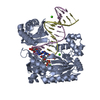


 PDBj
PDBj


 Assembly


 Mass: 40.078 Da / Num. of mol.: 3 / Source method: obtained synthetically / Formula: Ca
 Mass: 18.015 Da / Num. of mol.: 144 / Source method: isolated from a natural source / Formula: H2O
 Sample preparation
 / Beamline: 14-ID-B / Wavelength: 0.991 Å
 Processing