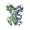
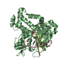
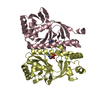
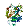
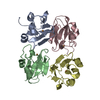
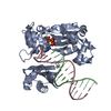
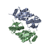
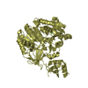
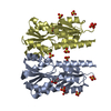
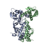
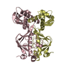
Catabolitegeneactivator / cAMP receptor protein / cAMP regulatory protein
Mass: 23672.439 Da / Num. of mol.: 6 Source method: isolated from a genetically manipulated source Source: (gene. exp.) Escherichia coli (E. coli) / Gene: crp, S4387, SF3376 / Plasmid: pET30a / Production host: Escherichia coli (E. coli) / Strain (production host): K12 / References: UniProt: P0ACK1, UniProt: P0ACJ8*PLUS
-
Experimental details
-
Experiment
Experiment
Method: X-RAY DIFFRACTION / Number of used crystals: 1
-
Sample preparation
Crystal
Density Matthews: 3.58 Å3/Da / Density % sol: 65.68 %
Crystal grow
Temperature: 298 K / Method: vapor diffusion / pH: 7.5 Details: Protein was crystallized after it was concentrated to ~25 mg/ml and seeded with apo D138L mutant CAP., pH 7.5, VAPOR DIFFUSION, temperature 298K
Resolution: 3.6→3.73 Å / Redundancy: 5.7 % / Mean I/σ(I) obs: 2.621
-
Processing
Software
Name
Version
Classification
HKL-2000
datacollection
DM
modelbuilding
REFMAC
5.4.0077
refinement
HKL-2000
datareduction
HKL-2000
datascaling
DM
phasing
Refinement
Method to determine structure: AVERAGING Starting model: cAMP Binding Domain of CAP Resolution: 3.59→48.06 Å / Cor.coef. Fo:Fc: 0.889 / Cor.coef. Fo:Fc free: 0.881 / SU B: 132.978 / SU ML: 0.837 / TLS residual ADP flag: LIKELY RESIDUAL / Cross valid method: THROUGHOUT / ESU R Free: 0.75 / Stereochemistry target values: MAXIMUM LIKELIHOOD / Details: HYDROGENS HAVE BEEN ADDED IN THE RIDING POSITIONS
Rfactor
Num. reflection
% reflection
Selection details
Rfree
0.31823
1217
5 %
RANDOM
Rwork
0.29492
-
-
-
obs
0.29608
22948
98.93 %
-
Solvent computation
Ion probe radii: 0.8 Å / Shrinkage radii: 0.8 Å / VDW probe radii: 1.2 Å / Solvent model: MASK
Displacement parameters
Biso mean: 29.723 Å2
Baniso -1
Baniso -2
Baniso -3
1-
3.56 Å2
1.78 Å2
0 Å2
2-
-
3.56 Å2
0 Å2
3-
-
-
-5.35 Å2
Refinement step
Cycle: LAST / Resolution: 3.59→48.06 Å
Protein
Nucleic acid
Ligand
Solvent
Total
Num. atoms
9456
0
0
0
9456
Refine LS restraints
Refine-ID
Type
Dev ideal
Dev ideal target
Number
X-RAY DIFFRACTION
r_bond_refined_d
0.004
0.022
9600
X-RAY DIFFRACTION
r_bond_other_d
X-RAY DIFFRACTION
r_angle_refined_deg
0.893
1.968
12924
X-RAY DIFFRACTION
r_angle_other_deg
X-RAY DIFFRACTION
r_dihedral_angle_1_deg
4.139
5
1182
X-RAY DIFFRACTION
r_dihedral_angle_2_deg
28.346
24.429
420
X-RAY DIFFRACTION
r_dihedral_angle_3_deg
15.369
15
1854
X-RAY DIFFRACTION
r_dihedral_angle_4_deg
13.491
15
60
X-RAY DIFFRACTION
r_chiral_restr
0.065
0.2
1488
X-RAY DIFFRACTION
r_gen_planes_refined
0.002
0.021
6966
X-RAY DIFFRACTION
r_gen_planes_other
X-RAY DIFFRACTION
r_nbd_refined
X-RAY DIFFRACTION
r_nbd_other
X-RAY DIFFRACTION
r_nbtor_refined
X-RAY DIFFRACTION
r_nbtor_other
X-RAY DIFFRACTION
r_xyhbond_nbd_refined
X-RAY DIFFRACTION
r_xyhbond_nbd_other
X-RAY DIFFRACTION
r_metal_ion_refined
X-RAY DIFFRACTION
r_metal_ion_other
X-RAY DIFFRACTION
r_symmetry_vdw_refined
X-RAY DIFFRACTION
r_symmetry_vdw_other
X-RAY DIFFRACTION
r_symmetry_hbond_refined
X-RAY DIFFRACTION
r_symmetry_hbond_other
X-RAY DIFFRACTION
r_symmetry_metal_ion_refined
X-RAY DIFFRACTION
r_symmetry_metal_ion_other
X-RAY DIFFRACTION
r_mcbond_it
0.103
1.5
5904
X-RAY DIFFRACTION
r_mcbond_other
X-RAY DIFFRACTION
r_mcangle_it
0.19
2
9522
X-RAY DIFFRACTION
r_scbond_it
0.164
3
3696
X-RAY DIFFRACTION
r_scangle_it
0.301
4.5
3402
X-RAY DIFFRACTION
r_rigid_bond_restr
X-RAY DIFFRACTION
r_sphericity_free
X-RAY DIFFRACTION
r_sphericity_bonded
LS refinement shell
Resolution: 3.594→3.687 Å / Total num. of bins used: 20
Rfactor
Num. reflection
% reflection
Rfree
0.357
91
-
Rwork
0.418
1573
-
obs
-
-
93.33 %
Refinement TLS params.
Method: refined / Refine-ID: X-RAY DIFFRACTION
ID
L11 (°2)
L12 (°2)
L13 (°2)
L22 (°2)
L23 (°2)
L33 (°2)
S11 (Å °)
S12 (Å °)
S13 (Å °)
S21 (Å °)
S22 (Å °)
S23 (Å °)
S31 (Å °)
S32 (Å °)
S33 (Å °)
T11 (Å2)
T12 (Å2)
T13 (Å2)
T22 (Å2)
T23 (Å2)
T33 (Å2)
Origin x (Å)
Origin y (Å)
Origin z (Å)
1
5.0524
3.6225
-1.6757
8.1234
-0.2766
7.9562
-0.0259
-0.0766
-0.0893
0.0332
-0.1921
0.9138
0.606
-0.8823
0.2179
1.6873
0.7506
0.1683
0.566
-0.045
1.4279
-28.073
2.832
44.944
2
3.1321
3.0503
-1.423
9.8741
0.9135
5.1871
-0.046
0.4693
-0.0816
-0.0215
0.0054
0.6109
0.1889
-0.5098
0.0406
1.3751
0.8571
-0.0243
0.9061
-0.0442
1.326
-14.981
-3.5
23.962
3
9.3531
-0.2831
-1.324
9.0106
3.9606
11.2802
-0.1948
-1.1381
-0.2916
0.8219
-0.4166
-0.747
0.9375
0.3322
0.6115
1.896
0.4185
-0.1064
1.1395
0.1861
0.7658
-24.863
-25.285
47.829
4
7.9239
-3.4038
-0.1596
4.1614
5.2515
12.255
-0.4655
-0.2482
-0.29
0.3269
0.3318
0.5999
0.9908
-0.6457
0.1337
1.9136
-0.0329
0.1762
1.1603
-0.0287
0.6663
-42.321
-25.74
30.424
5
4.9041
-1.6039
-1.249
9.5605
3.361
6.3316
0.432
0.5863
0.2093
-0.869
-0.6328
0.0405
-0.1865
-0.0795
0.2009
1.8362
1.2075
0.0981
0.8139
0.2199
1.22
22.212
-30.375
17.934
6
8.34
2.6792
-0.273
5.7294
0.6848
7.146
-0.4376
1.1043
-0.5378
-0.5433
0.2932
0.6564
0.4798
-0.7424
0.1445
1.8431
0.9307
0.0299
0.7868
-0.1936
1.3435
-3.155
-31.071
21.845
7
14.9492
3.4506
-6.546
5.7434
-6.2068
7.3239
-0.5488
0.652
0.9373
0.3339
0.6074
-0.3095
-0.0615
0.8633
-0.0586
1.3249
0.4362
0.0411
1.0528
-0.3839
1.0187
11.609
-44.321
-9.792
8
9.8675
2.7466
-3.513
4.2456
-3.9203
5.5055
0.7395
-0.6836
-0.4504
0.6911
-0.8057
0.226
1.5142
0.3434
0.0661
2.0851
0.5489
0.1316
1.5787
-0.264
1.123
1.849
-58.135
8.281
9
2.9837
0.9453
-1.3567
10.303
-3.9767
5.0468
0.0304
-0.1607
-0.3464
0.3521
-0.11
0.6515
-0.1563
-0.6697
0.0796
1.5772
0.8335
0.2234
0.8541
-0.1216
1.4837
-39.586
30.338
43.385
10
3.8307
0.5219
1.3922
5.2178
-0.3429
9.0752
0.0896
-0.503
-0.5029
0.8042
-0.1395
0.208
0.5198
-0.428
0.0499
2.0439
0.7339
0.1221
0.7464
0.1455
1.108
-33.289
47.914
60.797
11
2.5248
-3.5848
-2.3616
8.0449
6.4116
5.3746
-0.9196
-0.1132
-0.5325
-0.6075
-0.252
3.6703
-0.2247
-2.653
1.1717
2.8294
0.0685
-0.1015
2.1997
0.1289
2.5634
-46.125
18.745
73.115
12
2.4513
-2.6255
-2.1519
4.9979
2.7512
1.9803
-0.3485
-0.1829
0.2232
0.2924
0.6294
-0.6825
-0.8298
0.928
-0.281
2.9675
-0.1752
0.1761
2.004
0.1034
1.385
-22.399
24.266
71.722
Refinement TLS group
ID
Refine-ID
Refine TLS-ID
Auth asym-ID
Auth seq-ID
1
X-RAY DIFFRACTION
1
A
7 - 130
2
X-RAY DIFFRACTION
2
B
7 - 130
3
X-RAY DIFFRACTION
3
A
133 - 207
4
X-RAY DIFFRACTION
4
B
133 - 207
5
X-RAY DIFFRACTION
5
C
7 - 130
6
X-RAY DIFFRACTION
6
D
7 - 130
7
X-RAY DIFFRACTION
7
C
133 - 207
8
X-RAY DIFFRACTION
8
D
133 - 207
9
X-RAY DIFFRACTION
9
E
7 - 130
10
X-RAY DIFFRACTION
10
F
7 - 130
11
X-RAY DIFFRACTION
11
E
133 - 207
12
X-RAY DIFFRACTION
12
F
133 - 207
+
About Yorodumi
-
News
-
Feb 9, 2022. New format data for meta-information of EMDB entries
New format data for meta-information of EMDB entries
Version 3 of the EMDB header file is now the official format.
The previous official version 1.9 will be removed from the archive.
In the structure databanks used in Yorodumi, some data are registered as the other names, "COVID-19 virus" and "2019-nCoV". Here are the details of the virus and the list of structure data.
Jan 31, 2019. EMDB accession codes are about to change! (news from PDBe EMDB page)
EMDB accession codes are about to change! (news from PDBe EMDB page)
The allocation of 4 digits for EMDB accession codes will soon come to an end. Whilst these codes will remain in use, new EMDB accession codes will include an additional digit and will expand incrementally as the available range of codes is exhausted. The current 4-digit format prefixed with “EMD-” (i.e. EMD-XXXX) will advance to a 5-digit format (i.e. EMD-XXXXX), and so on. It is currently estimated that the 4-digit codes will be depleted around Spring 2019, at which point the 5-digit format will come into force.
The EM Navigator/Yorodumi systems omit the EMD- prefix.
Related info.:Q: What is EMD? / ID/Accession-code notation in Yorodumi/EM Navigator
Yorodumi is a browser for structure data from EMDB, PDB, SASBDB, etc.
This page is also the successor to EM Navigator detail page, and also detail information page/front-end page for Omokage search.
The word "yorodu" (or yorozu) is an old Japanese word meaning "ten thousand". "mi" (miru) is to see.
Related info.:EMDB / PDB / SASBDB / Comparison of 3 databanks / Yorodumi Search / Aug 31, 2016. New EM Navigator & Yorodumi / Yorodumi Papers / Jmol/JSmol / Function and homology information / Changes in new EM Navigator and Yorodumi
 Movie
Movie Controller


 Open data
 Basic information
 Components
 Keywords
 TRANSCRIPTION REGULATOR /
  Function and homology information
 Authors
 Citation
 Structure visualization
 Downloads & links
 Other downloads

 PDBj
PDBj


 Assembly

 Sample preparation
 / Beamline: 24-ID-C / Wavelength: 0.98 Å
 Processing