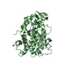
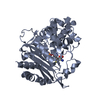
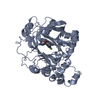
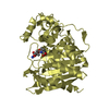
- PDB-6bxi: X-ray crystal structure of NDR1 kinase domain -
+
Open data
ID or keywords:
Loading...
-
Basic information
Entry
Database: PDB / ID: 6bxi
Title
X-ray crystal structure of NDR1 kinase domain
Components
Serine/threonine-protein kinase 38Serine/threonine-specific protein kinase
Keywords
TRANSFERASE / kinase fold with atypically long activation segment
Function / homology
Function and homology information
postsynapse organization / mitogen-activated protein kinase kinase kinase binding / RHOBTB1 GTPase cycle / RHOBTB2 GTPase cycle / negative regulation of MAP kinase activity / protein modification process / non-specific serine/threonine protein kinase / intracellular signal transduction / cadherin binding / protein phosphorylation ...postsynapse organization / mitogen-activated protein kinase kinase kinase binding / RHOBTB1 GTPase cycle / RHOBTB2 GTPase cycle / negative regulation of MAP kinase activity / protein modification process / non-specific serine/threonine protein kinase / intracellular signal transduction / cadherin binding / protein phosphorylation / protein serine kinase activity / protein serine/threonine kinase activity / glutamatergic synapse / magnesium ion binding / ATP binding / nucleus / cytosol / cytoplasm Similarity search - Function
Protein kinase, C-terminal / Protein kinase C terminal domain / Extension to Ser/Thr-type protein kinases / AGC-kinase, C-terminal / AGC-kinase C-terminal domain profile. / Serine/threonine-protein kinase, active site / Serine/Threonine protein kinases active-site signature. / Protein kinase domain / Serine/Threonine protein kinases, catalytic domain / Protein kinase, ATP binding site ...Protein kinase, C-terminal / Protein kinase C terminal domain / Extension to Ser/Thr-type protein kinases / AGC-kinase, C-terminal / AGC-kinase C-terminal domain profile. / Serine/threonine-protein kinase, active site / Serine/Threonine protein kinases active-site signature. / Protein kinase domain / Serine/Threonine protein kinases, catalytic domain / Protein kinase, ATP binding site / Protein kinases ATP-binding region signature. / Protein kinase domain profile. / Protein kinase domain / Protein kinase-like domain superfamily Similarity search - Domain/homology
Serine/threonine-proteinkinase38 / Serine/threonine-specific protein kinase / NDR1 protein kinase / Nuclear Dbf2-related kinase 1
Mass: 38753.465 Da / Num. of mol.: 2 Source method: isolated from a genetically manipulated source Source: (gene. exp.) Homo sapiens (human) / Gene: STK38, NDR1 / Production host: Escherichia coli (E. coli) References: UniProt: Q15208, non-specific serine/threonine protein kinase
In the structure databanks used in Yorodumi, some data are registered as the other names, "COVID-19 virus" and "2019-nCoV". Here are the details of the virus and the list of structure data.
Jan 31, 2019. EMDB accession codes are about to change! (news from PDBe EMDB page)
EMDB accession codes are about to change! (news from PDBe EMDB page)
The allocation of 4 digits for EMDB accession codes will soon come to an end. Whilst these codes will remain in use, new EMDB accession codes will include an additional digit and will expand incrementally as the available range of codes is exhausted. The current 4-digit format prefixed with “EMD-” (i.e. EMD-XXXX) will advance to a 5-digit format (i.e. EMD-XXXXX), and so on. It is currently estimated that the 4-digit codes will be depleted around Spring 2019, at which point the 5-digit format will come into force.
The EM Navigator/Yorodumi systems omit the EMD- prefix.
Related info.:Q: What is EMD? / ID/Accession-code notation in Yorodumi/EM Navigator
Yorodumi is a browser for structure data from EMDB, PDB, SASBDB, etc.
This page is also the successor to EM Navigator detail page, and also detail information page/front-end page for Omokage search.
The word "yorodu" (or yorozu) is an old Japanese word meaning "ten thousand". "mi" (miru) is to see.
Related info.:EMDB / PDB / SASBDB / Comparison of 3 databanks / Yorodumi Search / Aug 31, 2016. New EM Navigator & Yorodumi / Yorodumi Papers / Jmol/JSmol / Function and homology information / Changes in new EM Navigator and Yorodumi
 Movie
Movie Controller


 Open data
 Basic information
 Components
 Serine/threonine-specific protein kinase
  Keywords
 Function and homology information
 Authors
 Citation
 Structure visualization
 Downloads & links
 Other downloads
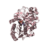


 PDBj
PDBj


 Assembly
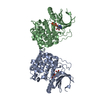

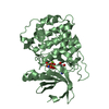


 Mass: 506.196 Da / Num. of mol.: 2 / Source method: obtained synthetically / Formula: C10H17N6O12P3 / Comment: AMP-PNP, energy-carrying molecule analogue*YM
 Mass: 24.305 Da / Num. of mol.: 2 / Source method: obtained synthetically / Formula: Mg
 Mass: 18.015 Da / Num. of mol.: 441 / Source method: isolated from a natural source / Formula: H2O
 Sample preparation
 / Beamline: 24-ID-C / Wavelength: 0.9792 Å
 Processing