 Movie
Movie Controller

[English] 日本語
 Yorodumi
- PDB-6rcg: Crystal structure of Casein kinase 1 delta (CK1 delta) complexed ... -
+ Open data

- Basic information
| Entry | Database: PDB / ID: 6rcg | ||||||
|---|---|---|---|---|---|---|---|
| Title | Crystal structure of Casein kinase 1 delta (CK1 delta) complexed with SR3029 inhibitor | ||||||
 Components Components | Casein kinase I isoform delta | ||||||
 Keywords Keywords |  TRANSFERASE / CSNK1D / CK1 delta / CK1d / kinase inhibitor / Structural Genomics / Structural Genomics Consortium / SGC TRANSFERASE / CSNK1D / CK1 delta / CK1d / kinase inhibitor / Structural Genomics / Structural Genomics Consortium / SGC | ||||||
| Function / homology |  Function and homology information Function and homology informationpositive regulation of non-canonical Wnt signaling pathway / protein localization to Golgi apparatus / COPII vesicle coating / midbrain dopaminergic neuron differentiation / microtubule nucleation / protein localization to cilium / tau-protein kinase / non-motile cilium assembly / protein localization to centrosome / COPII-mediated vesicle transport ...positive regulation of non-canonical Wnt signaling pathway / protein localization to Golgi apparatus / COPII vesicle coating / midbrain dopaminergic neuron differentiation / microtubule nucleation / protein localization to cilium / tau-protein kinase / non-motile cilium assembly / protein localization to centrosome / COPII-mediated vesicle transport / tau-protein kinase activity / Golgi organization / Major pathway of rRNA processing in the nucleolus and cytosol / spindle assembly / Loss of Nlp from mitotic centrosomes / Loss of proteins required for interphase microtubule organization from the centrosome / Recruitment of mitotic centrosome proteins and complexes / Recruitment of NuMA to mitotic centrosomes / Anchoring of the basal body to the plasma membrane / endoplasmic reticulum-Golgi intermediate compartment membrane / AURKA Activation by TPX2 / cellular response to nerve growth factor stimulus / ciliary basal body / circadian regulation of gene expression / spindle microtubule / regulation of circadian rhythm / Wnt signaling pathway / spindle / endocytosis / Regulation of PLK1 Activity at G2/M Transition / positive regulation of canonical Wnt signaling pathway / Circadian Clock / positive regulation of proteasomal ubiquitin-dependent protein catabolic process / non-specific serine/threonine protein kinase / protein kinase activity / cadherin binding / positive regulation of protein phosphorylation / protein phosphorylation / protein serine kinase activity / protein serine/threonine kinase activity / centrosome / perinuclear region of cytoplasm / Golgi apparatus / signal transduction / nucleoplasm / ATP binding / nucleus / plasma membrane / cytosolSimilarity search - Function | ||||||
| Biological species |  Homo sapiens (human) Homo sapiens (human) | ||||||
| Method | X-RAY DIFFRACTION / SYNCHROTRON / MOLECULAR REPLACEMENT / Resolution: 1.4 Å | ||||||
 Authors Authors | Chaikuad, A. / Arrowsmith, C.H. / Edwards, A.M. / Bountra, C. / Roush, W.R. / Knapp, S. / Structural Genomics Consortium (SGC) | ||||||
 Citation Citation | Journal: To Be Published Title: Crystal structure of Casein kinase 1 delta (CK1 delta) complexed with SR3029 inhibitor Authors: Chaikuad, A. / Arrowsmith, C.H. / Edwards, A.M. / Bountra, C. / Roush, W.R. / Knapp, S. / Structural Genomics Consortium (SGC) | ||||||
| History |
|
- Structure visualization
| Structure viewer | Molecule: MolmilJmol/JSmol |
|---|
- Downloads & links
-Download
| PDBx/mmCIF format | 6rcg.cif.gz | 146.7 KB | Display | PDBx/mmCIF format |
|---|---|---|---|---|
| PDB format | pdb6rcg.ent.gz | 114.1 KB | Display | PDB format |
| PDBx/mmJSON format | 6rcg.json.gz | Tree view | PDBx/mmJSON format | |
| Others |  Other downloads Other downloads |
-Validation report
| Arichive directory | https://data.pdbj.org/pub/pdb/validation_reports/rc/6rcgftp://data.pdbj.org/pub/pdb/validation_reports/rc/6rcg | HTTPS FTP |
|---|
-Related structure data
| Related structure data |  4hnfS S: Starting model for refinement |
|---|---|
| Similar structure data |


-Links
 PDBj
PDBj


- Assembly
| Deposited unit | 
| ||||||||
|---|---|---|---|---|---|---|---|---|---|
| 1 | 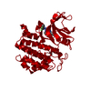
| ||||||||
| Unit cell |
|
-Components
| #1: Protein | Mass: 34424.805 Da / Num. of mol.: 1 Source method: isolated from a genetically manipulated source Source: (gene. exp.) Homo sapiens (human) / Gene: CSNK1D, HCKID / Plasmid: pNIC28-Bsa4 / Production host:  Escherichia coli (E. coli) / Strain (production host): BL21(DE3)-R3-pRARE2 Escherichia coli (E. coli) / Strain (production host): BL21(DE3)-R3-pRARE2References: UniProt: P48730, non-specific serine/threonine protein kinase, tau-protein kinase | ||||
|---|---|---|---|---|---|
| #2: Chemical | Ethylene glycol  Mass: 62.068 Da / Num. of mol.: 2 / Source method: obtained synthetically / Formula: C2H6O2 Mass: 62.068 Da / Num. of mol.: 2 / Source method: obtained synthetically / Formula: C2H6O2#3: Chemical | ChemComp-K0E / ~{ |   Mass: 480.445 Da / Num. of mol.: 1 / Source method: obtained synthetically / Formula: C23H19F3N8O Mass: 480.445 Da / Num. of mol.: 1 / Source method: obtained synthetically / Formula: C23H19F3N8O#4: Water | ChemComp-HOH / | Water Mass: 18.015 Da / Num. of mol.: 381 / Source method: isolated from a natural source / Formula: H2O Mass: 18.015 Da / Num. of mol.: 381 / Source method: isolated from a natural source / Formula: H2O |
-Experimental details
-Experiment
| Experiment | Method: X-RAY DIFFRACTION / Number of used crystals: 1 |
|---|
- Sample preparation
| Crystal | Density Matthews: 2.27 Å3/Da / Density % sol: 45.74 % |
|---|---|
| Crystal grow | Temperature: 277.15 K / Method: vapor diffusion, sitting drop / Details: 20% PEG3350, 0.1M succinic acid |
-Data collection
| Diffraction | Mean temperature: 100 K / Serial crystal experiment: N | ||||||||||||||||||||||||||||||||||||||||||||||||||||||||||||||||||||||||||||||||||||||||||||||||||||||||||||||
|---|---|---|---|---|---|---|---|---|---|---|---|---|---|---|---|---|---|---|---|---|---|---|---|---|---|---|---|---|---|---|---|---|---|---|---|---|---|---|---|---|---|---|---|---|---|---|---|---|---|---|---|---|---|---|---|---|---|---|---|---|---|---|---|---|---|---|---|---|---|---|---|---|---|---|---|---|---|---|---|---|---|---|---|---|---|---|---|---|---|---|---|---|---|---|---|---|---|---|---|---|---|---|---|---|---|---|---|---|---|---|---|
| Diffraction source | Source: SYNCHROTRON / Site: Diamond  / Beamline: I04-1 / Wavelength: 0.92 Å / Beamline: I04-1 / Wavelength: 0.92 Å | ||||||||||||||||||||||||||||||||||||||||||||||||||||||||||||||||||||||||||||||||||||||||||||||||||||||||||||||
| Detector | Type: DECTRIS PILATUS3 2M / Detector: PIXEL / Date: Jul 3, 2013 | ||||||||||||||||||||||||||||||||||||||||||||||||||||||||||||||||||||||||||||||||||||||||||||||||||||||||||||||
| Radiation | Protocol: SINGLE WAVELENGTH / Monochromatic (M) / Laue (L): M / Scattering type: x-ray | ||||||||||||||||||||||||||||||||||||||||||||||||||||||||||||||||||||||||||||||||||||||||||||||||||||||||||||||
| Radiation wavelength | Wavelength: 0.92 Å / Relative weight: 1 | ||||||||||||||||||||||||||||||||||||||||||||||||||||||||||||||||||||||||||||||||||||||||||||||||||||||||||||||
| Reflection | Resolution: 1.4→66.209 Å / Num. obs: 58592 / % possible obs: 96.7 % / Redundancy: 3.6 % / CC1/2: 0.998 / Rpim(I) all: 0.031 / Rrim(I) all: 0.06 / Rsym value: 0.051 / Net I/av σ(I): 6.4 / Net I/σ(I): 12 | ||||||||||||||||||||||||||||||||||||||||||||||||||||||||||||||||||||||||||||||||||||||||||||||||||||||||||||||
| Reflection shell | Diffraction-ID: 1
|
- Processing
| Software |
| ||||||||||||||||||||||||||||||||||||||||||||||||||||||||||||||||||||||
|---|---|---|---|---|---|---|---|---|---|---|---|---|---|---|---|---|---|---|---|---|---|---|---|---|---|---|---|---|---|---|---|---|---|---|---|---|---|---|---|---|---|---|---|---|---|---|---|---|---|---|---|---|---|---|---|---|---|---|---|---|---|---|---|---|---|---|---|---|---|---|---|
| Refinement | Method to determine structure: MOLECULAR REPLACEMENT Starting model: 4HNF Resolution: 1.4→29.63 Å / Cor.coef. Fo:Fc: 0.98 / Cor.coef. Fo:Fc free: 0.968 / SU B: 2.202 / SU ML: 0.043 / SU R Cruickshank DPI: 0.0621 / Cross valid method: THROUGHOUT / σ(F): 0 / ESU R: 0.062 / ESU R Free: 0.061 Details: HYDROGENS HAVE BEEN ADDED IN THE RIDING POSITIONS U VALUES : REFINED INDIVIDUALLY
| ||||||||||||||||||||||||||||||||||||||||||||||||||||||||||||||||||||||
| Solvent computation | Ion probe radii: 0.8 Å / Shrinkage radii: 0.8 Å / VDW probe radii: 1.2 Å | ||||||||||||||||||||||||||||||||||||||||||||||||||||||||||||||||||||||
| Displacement parameters | Biso max: 128.45 Å2 / Biso mean: 23.78 Å2 / Biso min: 10.07 Å2
| ||||||||||||||||||||||||||||||||||||||||||||||||||||||||||||||||||||||
| Refinement step | Cycle: final / Resolution: 1.4→29.63 Å
| ||||||||||||||||||||||||||||||||||||||||||||||||||||||||||||||||||||||
| Refine LS restraints |
| ||||||||||||||||||||||||||||||||||||||||||||||||||||||||||||||||||||||
| LS refinement shell | Resolution: 1.4→1.436 Å / Rfactor Rfree error: 0 / Total num. of bins used: 20
|
