 Movie
Movie Controller


+ Open data

- Basic information
| Entry | Database: PDB / ID: 6b9d | ||||||
|---|---|---|---|---|---|---|---|
| Title | Human ATL1 mutant - R77A bound to GDP | ||||||
 Components Components | Atlastin-1 | ||||||
 Keywords Keywords | HYDROLASE / GTPase / dynamin related protein / hydrolysis-deficient | ||||||
| Function / homology |  Function and homology information Function and homology informationendoplasmic reticulum tubular network membrane organization / endoplasmic reticulum tubular network membrane / Golgi cis cisterna / endoplasmic reticulum tubular network / endoplasmic reticulum organization / axonogenesis / Hydrolases; Acting on acid anhydrides; Acting on GTP to facilitate cellular and subcellular movement / protein homooligomerization / axon / Golgi membrane ...endoplasmic reticulum tubular network membrane organization / endoplasmic reticulum tubular network membrane / Golgi cis cisterna / endoplasmic reticulum tubular network / endoplasmic reticulum organization / axonogenesis / Hydrolases; Acting on acid anhydrides; Acting on GTP to facilitate cellular and subcellular movement / protein homooligomerization / axon / Golgi membrane / GTPase activity / endoplasmic reticulum membrane / GTP binding / Golgi apparatus / endoplasmic reticulum / membrane / identical protein bindingSimilarity search - Function | ||||||
| Biological species |  Homo sapiens (human) Homo sapiens (human) | ||||||
| Method | X-RAY DIFFRACTION / SYNCHROTRON / MOLECULAR REPLACEMENT / Resolution: 1.95 Å | ||||||
 Authors Authors | O'Donnell, J.P. / Sondermann, H. | ||||||
| Funding support |  United States, 1items United States, 1items
| ||||||
 Citation Citation | Journal: J. Biol. Chem. / Year: 2018 Title: A hereditary spastic paraplegia-associated atlastin variant exhibits defective allosteric coupling in the catalytic core. Authors: O'Donnell, J.P. / Byrnes, L.J. / Cooley, R.B. / Sondermann, H. | ||||||
| History |
|
- Structure visualization
| Structure viewer | Molecule: MolmilJmol/JSmol |
|---|
- Downloads & links
-Download
| PDBx/mmCIF format | 6b9d.cif.gz | 321 KB | Display | PDBx/mmCIF format |
|---|---|---|---|---|
| PDB format | pdb6b9d.ent.gz | 257.7 KB | Display | PDB format |
| PDBx/mmJSON format | 6b9d.json.gz | Tree view | PDBx/mmJSON format | |
| Others |  Other downloads Other downloads |
-Validation report
| Arichive directory | https://data.pdbj.org/pub/pdb/validation_reports/b9/6b9dftp://data.pdbj.org/pub/pdb/validation_reports/b9/6b9d | HTTPS FTP |
|---|
-Related structure data
| Related structure data |  6b9eC  6b9fC  6b9gC  3q5eS S: Starting model for refinement C: citing same article ( |
|---|---|
| Similar structure data |


-Links
 PDBj
PDBj- Assembly
| Deposited unit | 
| ||||||||
|---|---|---|---|---|---|---|---|---|---|
| 1 | 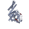
| ||||||||
| 2 | 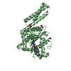
| ||||||||
| Unit cell |
|
-Components
| #1: Protein | / Brain-specific GTP-binding protein / GTP-binding protein 3 / hGBP3 / Guanine nucleotide-binding ...Brain-specific GTP-binding protein / GTP-binding protein 3 / hGBP3 / Guanine nucleotide-binding protein 3 / Spastic paraplegia 3 protein A Mass: 52502.340 Da / Num. of mol.: 2 / Fragment: residues 1-446 / Mutation: R77A Source method: isolated from a genetically manipulated source Source: (gene. exp.) Homo sapiens (human) / Gene: ATL1, GBP3, SPG3A / Production host:  Escherichia coli (E. coli) Escherichia coli (E. coli)References: UniProt: Q8WXF7, Hydrolases; Acting on acid anhydrides; Acting on GTP to facilitate cellular and subcellular movement#2: Chemical | Guanosine diphosphate  Type: RNA linking / Mass: 443.201 Da / Num. of mol.: 2 / Source method: obtained synthetically / Formula: C10H15N5O11P2 / Feature type: SUBJECT OF INVESTIGATION / Comment: GDP, energy-carrying molecule*YM Type: RNA linking / Mass: 443.201 Da / Num. of mol.: 2 / Source method: obtained synthetically / Formula: C10H15N5O11P2 / Feature type: SUBJECT OF INVESTIGATION / Comment: GDP, energy-carrying molecule*YM#3: Chemical |   Mass: 24.305 Da / Num. of mol.: 2 / Source method: obtained synthetically / Formula: Mg / Feature type: SUBJECT OF INVESTIGATION Mass: 24.305 Da / Num. of mol.: 2 / Source method: obtained synthetically / Formula: Mg / Feature type: SUBJECT OF INVESTIGATION#4: Chemical | ChemComp-GOL / Glycerol  Mass: 92.094 Da / Num. of mol.: 6 / Source method: obtained synthetically / Formula: C3H8O3 Mass: 92.094 Da / Num. of mol.: 6 / Source method: obtained synthetically / Formula: C3H8O3#5: Water | ChemComp-HOH / | Water Mass: 18.015 Da / Num. of mol.: 281 / Source method: isolated from a natural source / Formula: H2O Mass: 18.015 Da / Num. of mol.: 281 / Source method: isolated from a natural source / Formula: H2O |
|---|
-Experimental details
-Experiment
| Experiment | Method: X-RAY DIFFRACTION / Number of used crystals: 1 |
|---|
- Sample preparation
| Crystal | Density Matthews: 2.27 Å3/Da / Density % sol: 45.83 % |
|---|---|
| Crystal grow | Temperature: 293.15 K / Method: vapor diffusion, hanging drop Details: 0.2 M sodium malonate pH 6, 20% PEG3350, 1 mM GDP, 2 mM MgCl2 |
-Data collection
| Diffraction | Mean temperature: 100 K |
|---|---|
| Diffraction source | Source: SYNCHROTRON / Site: CHESS / Beamline: A1 / Wavelength: 0.976 Å |
| Detector | Type: ADSC QUANTUM 210 / Detector: CCD / Date: Jun 7, 2012 |
| Radiation | Protocol: SINGLE WAVELENGTH / Monochromatic (M) / Laue (L): M / Scattering type: x-ray |
| Radiation wavelength | Wavelength: 0.976 Å / Relative weight: 1 |
| Reflection | Resolution: 1.95→42.36 Å / Num. obs: 63315 / % possible obs: 96.4 % / Redundancy: 2.4 % / CC1/2: 0.991 / Rmerge(I) obs: 0.079 / Rrim(I) all: 0.103 / Net I/σ(I): 7.1 |
- Processing
| Software |
| |||||||||||||||||||||||||||||||||||||||||||||||||||||||||||||||||||||||||||||||||||||||||||||||||||||||||||||||||||||||||||||||||||||||||||||||||||||||||||||||||||||||||||||||
|---|---|---|---|---|---|---|---|---|---|---|---|---|---|---|---|---|---|---|---|---|---|---|---|---|---|---|---|---|---|---|---|---|---|---|---|---|---|---|---|---|---|---|---|---|---|---|---|---|---|---|---|---|---|---|---|---|---|---|---|---|---|---|---|---|---|---|---|---|---|---|---|---|---|---|---|---|---|---|---|---|---|---|---|---|---|---|---|---|---|---|---|---|---|---|---|---|---|---|---|---|---|---|---|---|---|---|---|---|---|---|---|---|---|---|---|---|---|---|---|---|---|---|---|---|---|---|---|---|---|---|---|---|---|---|---|---|---|---|---|---|---|---|---|---|---|---|---|---|---|---|---|---|---|---|---|---|---|---|---|---|---|---|---|---|---|---|---|---|---|---|---|---|---|---|---|---|
| Refinement | Method to determine structure: MOLECULAR REPLACEMENT Starting model: 3Q5E Resolution: 1.95→25.18 Å / SU ML: 0.25 / Cross valid method: FREE R-VALUE / σ(F): 1.96 / Phase error: 29.82
| |||||||||||||||||||||||||||||||||||||||||||||||||||||||||||||||||||||||||||||||||||||||||||||||||||||||||||||||||||||||||||||||||||||||||||||||||||||||||||||||||||||||||||||||
| Solvent computation | Shrinkage radii: 0.9 Å / VDW probe radii: 1.11 Å | |||||||||||||||||||||||||||||||||||||||||||||||||||||||||||||||||||||||||||||||||||||||||||||||||||||||||||||||||||||||||||||||||||||||||||||||||||||||||||||||||||||||||||||||
| Refinement step | Cycle: LAST / Resolution: 1.95→25.18 Å
| |||||||||||||||||||||||||||||||||||||||||||||||||||||||||||||||||||||||||||||||||||||||||||||||||||||||||||||||||||||||||||||||||||||||||||||||||||||||||||||||||||||||||||||||
| Refine LS restraints |
| |||||||||||||||||||||||||||||||||||||||||||||||||||||||||||||||||||||||||||||||||||||||||||||||||||||||||||||||||||||||||||||||||||||||||||||||||||||||||||||||||||||||||||||||
| LS refinement shell |
| |||||||||||||||||||||||||||||||||||||||||||||||||||||||||||||||||||||||||||||||||||||||||||||||||||||||||||||||||||||||||||||||||||||||||||||||||||||||||||||||||||||||||||||||
| Refinement TLS params. | Method: refined / Refine-ID: X-RAY DIFFRACTION
| |||||||||||||||||||||||||||||||||||||||||||||||||||||||||||||||||||||||||||||||||||||||||||||||||||||||||||||||||||||||||||||||||||||||||||||||||||||||||||||||||||||||||||||||
| Refinement TLS group |
|
