 Movie
Movie Controller

[English] 日本語
 Yorodumi
- PDB-3qof: Crystal structure of the cytosolic domain of human atlastin-1 in ... -
+ Open data

- Basic information
| Entry | Database: PDB / ID: 3qof | ||||||
|---|---|---|---|---|---|---|---|
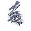
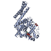
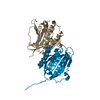
| Title | Crystal structure of the cytosolic domain of human atlastin-1 in complex with GDP, orthorhombic form | ||||||
 Components Components | Atlastin-1 | ||||||
 Keywords Keywords | HYDROLASE / GTPase / homotypic fusion / fusion of ER membranes / GDP / GTP / endoplasmic reticulum | ||||||
| Function / homology |  Function and homology information Function and homology informationendoplasmic reticulum tubular network membrane organization / endoplasmic reticulum tubular network membrane / Golgi cis cisterna / endoplasmic reticulum tubular network / endoplasmic reticulum organization / axonogenesis / Hydrolases; Acting on acid anhydrides; Acting on GTP to facilitate cellular and subcellular movement / protein homooligomerization / axon / Golgi membrane ...endoplasmic reticulum tubular network membrane organization / endoplasmic reticulum tubular network membrane / Golgi cis cisterna / endoplasmic reticulum tubular network / endoplasmic reticulum organization / axonogenesis / Hydrolases; Acting on acid anhydrides; Acting on GTP to facilitate cellular and subcellular movement / protein homooligomerization / axon / Golgi membrane / GTPase activity / endoplasmic reticulum membrane / GTP binding / Golgi apparatus / endoplasmic reticulum / membrane / identical protein bindingSimilarity search - Function | ||||||
| Biological species |  Homo sapiens (human) Homo sapiens (human) | ||||||
| Method | X-RAY DIFFRACTION / SYNCHROTRON / MOLECULAR REPLACEMENT / Resolution: 2.802 Å | ||||||
 Authors Authors | Liu, X. | ||||||
 Citation Citation | Journal: Proc.Natl.Acad.Sci.USA / Year: 2011 Title: Structures of the atlastin GTPase provide insight into homotypic fusion of endoplasmic reticulum membranes. Authors: Bian, X. / Klemm, R.W. / Liu, T.Y. / Zhang, M. / Sun, S. / Sui, X. / Liu, X. / Rapoport, T.A. / Hu, J. | ||||||
| History |
|
- Structure visualization
| Structure viewer | Molecule: MolmilJmol/JSmol |
|---|
- Downloads & links
-Download
| PDBx/mmCIF format | 3qof.cif.gz | 329.3 KB | Display | PDBx/mmCIF format |
|---|---|---|---|---|
| PDB format | pdb3qof.ent.gz | 259.7 KB | Display | PDB format |
| PDBx/mmJSON format | 3qof.json.gz | Tree view | PDBx/mmJSON format | |
| Others |  Other downloads Other downloads |
-Validation report
| Arichive directory | https://data.pdbj.org/pub/pdb/validation_reports/qo/3qofftp://data.pdbj.org/pub/pdb/validation_reports/qo/3qof | HTTPS FTP |
|---|
-Related structure data
| Related structure data |  3qnuSC S: Starting model for refinement C: citing same article ( |
|---|---|
| Similar structure data |


-Links
 PDBj
PDBj- Assembly
| Deposited unit | 
| ||||||||
|---|---|---|---|---|---|---|---|---|---|
| 1 | 
| ||||||||
| 2 | 
| ||||||||
| 3 | 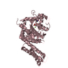
| ||||||||
| 4 | 
| ||||||||
| Unit cell |
|
-Components
| #1: Protein | / Brain-specific GTP-binding protein / GTP-binding protein 3 / GBP-3 / hGBP3 / Guanine nucleotide- ...Brain-specific GTP-binding protein / GTP-binding protein 3 / GBP-3 / hGBP3 / Guanine nucleotide-binding protein 3 / Spastic paraplegia 3 protein A Mass: 52718.609 Da / Num. of mol.: 4 / Fragment: atlastin ecto-domain (UNP RESIDUES 18-447) Source method: isolated from a genetically manipulated source Source: (gene. exp.) Homo sapiens (human) / Gene: ATL1, GBP3, SPG3A / Plasmid: pet30 / Production host:  Escherichia coli (E. coli) / Strain (production host): BL21(DE3) Escherichia coli (E. coli) / Strain (production host): BL21(DE3)References: UniProt: Q8WXF7, Hydrolases; Acting on acid anhydrides; Acting on GTP to facilitate cellular and subcellular movement#2: Chemical | ChemComp-GDP / Guanosine diphosphate  Type: RNA linking / Mass: 443.201 Da / Num. of mol.: 4 / Source method: obtained synthetically / Formula: C10H15N5O11P2 / Comment: GDP, energy-carrying molecule*YM Type: RNA linking / Mass: 443.201 Da / Num. of mol.: 4 / Source method: obtained synthetically / Formula: C10H15N5O11P2 / Comment: GDP, energy-carrying molecule*YM#3: Chemical | ChemComp-MG /   Mass: 24.305 Da / Num. of mol.: 4 / Source method: obtained synthetically / Formula: Mg Mass: 24.305 Da / Num. of mol.: 4 / Source method: obtained synthetically / Formula: Mg#4: Water | ChemComp-HOH / | Water Mass: 18.015 Da / Num. of mol.: 126 / Source method: isolated from a natural source / Formula: H2O Mass: 18.015 Da / Num. of mol.: 126 / Source method: isolated from a natural source / Formula: H2O |
|---|
-Experimental details
-Experiment
| Experiment | Method: X-RAY DIFFRACTION / Number of used crystals: 1 |
|---|
- Sample preparation
| Crystal | Density Matthews: 2.92 Å3/Da / Density % sol: 57.89 % |
|---|---|
| Crystal grow | Temperature: 293 K / Method: vapor diffusion, hanging drop / pH: 8.3 Details: 0.1M Tris-HCl, pH8.3, 25%(w/v) PEG 3350, 0.1M (NH4)2HPO4, VAPOR DIFFUSION, HANGING DROP, temperature 293K |
-Data collection
| Diffraction | Mean temperature: 100 K | |||||||||||||||||||||||||||||||||||||||||||||||||
|---|---|---|---|---|---|---|---|---|---|---|---|---|---|---|---|---|---|---|---|---|---|---|---|---|---|---|---|---|---|---|---|---|---|---|---|---|---|---|---|---|---|---|---|---|---|---|---|---|---|---|
| Diffraction source | Source: SYNCHROTRON / Site: SSRF  / Beamline: BL17U / Wavelength: 0.9793 Å / Beamline: BL17U / Wavelength: 0.9793 Å | |||||||||||||||||||||||||||||||||||||||||||||||||
| Detector | Type: MAR CCD 165 mm / Detector: CCD / Date: Jul 14, 2010 / Details: mirrors | |||||||||||||||||||||||||||||||||||||||||||||||||
| Radiation | Monochromator: graphite / Protocol: SINGLE WAVELENGTH / Monochromatic (M) / Laue (L): M / Scattering type: x-ray | |||||||||||||||||||||||||||||||||||||||||||||||||
| Radiation wavelength | Wavelength: 0.9793 Å / Relative weight: 1 | |||||||||||||||||||||||||||||||||||||||||||||||||
| Reflection | Resolution: 2.8→48.751 Å / Num. all: 61398 / Num. obs: 60656 / % possible obs: 98.79 % / Observed criterion σ(F): 1 / Observed criterion σ(I): 1 / Redundancy: 7.2 % / Biso Wilson estimate: 57.4 Å2 / Rmerge(I) obs: 0.078 / Rsym value: 0.078 / Net I/σ(I): 12.1 | |||||||||||||||||||||||||||||||||||||||||||||||||
| Reflection shell |
|
- Processing
| Software |
| ||||||||||||||||||||||||||||||||||||||||||||||||||||||||||||||||||||||||||||||||||||||||||||||||||||||||||||||||||||||||||||||||||||||||||||||||||||||||||||||||||||||||||||||||||||||||
|---|---|---|---|---|---|---|---|---|---|---|---|---|---|---|---|---|---|---|---|---|---|---|---|---|---|---|---|---|---|---|---|---|---|---|---|---|---|---|---|---|---|---|---|---|---|---|---|---|---|---|---|---|---|---|---|---|---|---|---|---|---|---|---|---|---|---|---|---|---|---|---|---|---|---|---|---|---|---|---|---|---|---|---|---|---|---|---|---|---|---|---|---|---|---|---|---|---|---|---|---|---|---|---|---|---|---|---|---|---|---|---|---|---|---|---|---|---|---|---|---|---|---|---|---|---|---|---|---|---|---|---|---|---|---|---|---|---|---|---|---|---|---|---|---|---|---|---|---|---|---|---|---|---|---|---|---|---|---|---|---|---|---|---|---|---|---|---|---|---|---|---|---|---|---|---|---|---|---|---|---|---|---|---|---|---|
| Refinement | Method to determine structure: MOLECULAR REPLACEMENT Starting model: PDB ENTRY 3QNU Resolution: 2.802→40 Å / SU ML: 0.4 / Isotropic thermal model: isotropic / Cross valid method: THROUGHOUT / σ(F): 1.34 / σ(I): 1 / Stereochemistry target values: ML / Details: Used weighted full matrix least squares procedure
| ||||||||||||||||||||||||||||||||||||||||||||||||||||||||||||||||||||||||||||||||||||||||||||||||||||||||||||||||||||||||||||||||||||||||||||||||||||||||||||||||||||||||||||||||||||||||
| Solvent computation | Shrinkage radii: 0.9 Å / VDW probe radii: 1.11 Å / Solvent model: FLAT BULK SOLVENT MODEL / Bsol: 49.413 Å2 / ksol: 0.302 e/Å3 | ||||||||||||||||||||||||||||||||||||||||||||||||||||||||||||||||||||||||||||||||||||||||||||||||||||||||||||||||||||||||||||||||||||||||||||||||||||||||||||||||||||||||||||||||||||||||
| Displacement parameters | Biso mean: 65.8 Å2
| ||||||||||||||||||||||||||||||||||||||||||||||||||||||||||||||||||||||||||||||||||||||||||||||||||||||||||||||||||||||||||||||||||||||||||||||||||||||||||||||||||||||||||||||||||||||||
| Refine analyze |
| ||||||||||||||||||||||||||||||||||||||||||||||||||||||||||||||||||||||||||||||||||||||||||||||||||||||||||||||||||||||||||||||||||||||||||||||||||||||||||||||||||||||||||||||||||||||||
| Refinement step | Cycle: LAST / Resolution: 2.802→40 Å
| ||||||||||||||||||||||||||||||||||||||||||||||||||||||||||||||||||||||||||||||||||||||||||||||||||||||||||||||||||||||||||||||||||||||||||||||||||||||||||||||||||||||||||||||||||||||||
| Refine LS restraints |
| ||||||||||||||||||||||||||||||||||||||||||||||||||||||||||||||||||||||||||||||||||||||||||||||||||||||||||||||||||||||||||||||||||||||||||||||||||||||||||||||||||||||||||||||||||||||||
| LS refinement shell | Refine-ID: X-RAY DIFFRACTION / Total num. of bins used: 22
|
