 Movie
Movie Controller


+ Open data

- Basic information
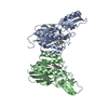
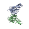
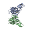
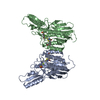
| Entry | Database: PDB / ID: 6asc | ||||||
|---|---|---|---|---|---|---|---|
| Title | Mre11 dimer in complex with Endonuclease inhibitor PFM04 | ||||||
 Components Components | Nuclease SbcCD subunit D | ||||||
 Keywords Keywords | hydrolase/hydrolase inhibitor / DNA REPAIR MRE11 THERMOPHILIC NUCLEASE DNA DOUBLE-STRAND BREAK REPAIR HYDROLASE / hydrolase-hydrolase inhibitor complex | ||||||
| Function / homology |  Function and homology information Function and homology informationDNA exonuclease activity / 3'-5' exonuclease activity / DNA endonuclease activity / double-strand break repair / DNA recombination /  DNA replication / Hydrolases; Acting on ester bonds / DNA repair / DNA binding / metal ion binding DNA replication / Hydrolases; Acting on ester bonds / DNA repair / DNA binding / metal ion bindingSimilarity search - Function | ||||||
| Biological species |   Thermotoga maritima (bacteria) Thermotoga maritima (bacteria) | ||||||
| Method | X-RAY DIFFRACTION / SYNCHROTRON / Resolution: 2.15 Å | ||||||
 Authors Authors | Moiani, D. / Arvai, A.S. / Tainer, J.A. | ||||||
 Citation Citation | Journal: Meth. Enzymol. / Year: 2018 Title: Targeting Allostery with Avatars to Design Inhibitors Assessed by Cell Activity: Dissecting MRE11 Endo- and Exonuclease Activities. Authors: Moiani, D. / Ronato, D.A. / Brosey, C.A. / Arvai, A.S. / Syed, A. / Masson, J.Y. / Petricci, E. / Tainer, J.A. | ||||||
| History |
|
- Structure visualization
| Structure viewer | Molecule: MolmilJmol/JSmol |
|---|
- Downloads & links
-Download
| PDBx/mmCIF format | 6asc.cif.gz | 276.5 KB | Display | PDBx/mmCIF format |
|---|---|---|---|---|
| PDB format | pdb6asc.ent.gz | 223.9 KB | Display | PDB format |
| PDBx/mmJSON format | 6asc.json.gz | Tree view | PDBx/mmJSON format | |
| Others |  Other downloads Other downloads |
-Validation report
| Arichive directory | https://data.pdbj.org/pub/pdb/validation_reports/as/6ascftp://data.pdbj.org/pub/pdb/validation_reports/as/6asc | HTTPS FTP |
|---|
-Related structure data
| Similar structure data |
|---|


-Links
 PDBj
PDBj
- Assembly
| Deposited unit | 
| ||||||||
|---|---|---|---|---|---|---|---|---|---|
| 1 |
| ||||||||
| Unit cell |
|
-Components
| #1: Protein | Mass: 38575.215 Da / Num. of mol.: 2 Source method: isolated from a genetically manipulated source Source: (gene. exp.) Thermotoga maritima (bacteria) / Strain: ATCC 43589 / MSB8 / DSM 3109 / JCM 10099 / Gene: sbcD, TM_1635 / Production host: Escherichia coli (E. coli) / References: UniProt: Q9X1X0#2: Chemical | ChemComp-MN /   Mass: 54.938 Da / Num. of mol.: 4 / Source method: isolated from a natural source / Formula: Mn Mass: 54.938 Da / Num. of mol.: 4 / Source method: isolated from a natural source / Formula: Mn#3: Chemical |   Mass: 293.404 Da / Num. of mol.: 2 / Source method: obtained synthetically / Formula: C14H15NO2S2 / Feature type: SUBJECT OF INVESTIGATION Mass: 293.404 Da / Num. of mol.: 2 / Source method: obtained synthetically / Formula: C14H15NO2S2 / Feature type: SUBJECT OF INVESTIGATION#4: Chemical | ChemComp-EDO / | Ethylene glycol  Mass: 62.068 Da / Num. of mol.: 1 / Source method: obtained synthetically / Formula: C2H6O2 Mass: 62.068 Da / Num. of mol.: 1 / Source method: obtained synthetically / Formula: C2H6O2#5: Water | ChemComp-HOH / | Water Mass: 18.015 Da / Num. of mol.: 142 / Source method: isolated from a natural source / Formula: H2O Mass: 18.015 Da / Num. of mol.: 142 / Source method: isolated from a natural source / Formula: H2O |
|---|
-Experimental details
-Experiment
| Experiment | Method: X-RAY DIFFRACTION / Number of used crystals: 1 |
|---|
- Sample preparation
| Crystal | Density Matthews: 2.87 Å3/Da / Density % sol: 57.13 % |
|---|---|
| Crystal grow | Temperature: 291 K / Method: vapor diffusion, hanging drop Details: We added 5mM PFM04 (dissolved in DMSO) to 10 mg/ml TmMre11. The mother liquor was 10% PEG3350, 20 mM CaCl2, 0.1 M Mes pH 6.5. This was mixed 1:1 with the protein:inhibitor complex and set up ...Details: We added 5mM PFM04 (dissolved in DMSO) to 10 mg/ml TmMre11. The mother liquor was 10% PEG3350, 20 mM CaCl2, 0.1 M Mes pH 6.5. This was mixed 1:1 with the protein:inhibitor complex and set up as hanging drop vapor diffusion. For cryoprotection, 20% ethylene glycol was added to the mother liquor. |
-Data collection
| Diffraction | Mean temperature: 100 K |
|---|---|
| Diffraction source | Source: SYNCHROTRON / Site: ALS  / Beamline: 12.3.1 / Wavelength: 1.11583 Å / Beamline: 12.3.1 / Wavelength: 1.11583 Å |
| Detector | Type: ADSC QUANTUM 315r / Detector: CCD / Date: Dec 2, 2011 |
| Radiation | Protocol: SINGLE WAVELENGTH / Monochromatic (M) / Laue (L): M / Scattering type: x-ray |
| Radiation wavelength | Wavelength: 1.11583 Å / Relative weight: 1 |
| Reflection | Resolution: 2.15→50 Å / Num. obs: 43590 / % possible obs: 94.2 % / Redundancy: 4.8 % / Net I/σ(I): 46.71 |
- Processing
| Software |
| |||||||||||||||||||||||||||||||||||||||||||||||||||||||||||||||||||||||||||||||||||||||||||||||||||||||||||||||||||||||||||||||||||||||||||||||||||||||||||||||||||||||||||||||
|---|---|---|---|---|---|---|---|---|---|---|---|---|---|---|---|---|---|---|---|---|---|---|---|---|---|---|---|---|---|---|---|---|---|---|---|---|---|---|---|---|---|---|---|---|---|---|---|---|---|---|---|---|---|---|---|---|---|---|---|---|---|---|---|---|---|---|---|---|---|---|---|---|---|---|---|---|---|---|---|---|---|---|---|---|---|---|---|---|---|---|---|---|---|---|---|---|---|---|---|---|---|---|---|---|---|---|---|---|---|---|---|---|---|---|---|---|---|---|---|---|---|---|---|---|---|---|---|---|---|---|---|---|---|---|---|---|---|---|---|---|---|---|---|---|---|---|---|---|---|---|---|---|---|---|---|---|---|---|---|---|---|---|---|---|---|---|---|---|---|---|---|---|---|---|---|---|
| Refinement | Resolution: 2.15→46.038 Å / SU ML: 0.27 / Cross valid method: FREE R-VALUE / σ(F): 0 / Phase error: 29.73 / Stereochemistry target values: ML
| |||||||||||||||||||||||||||||||||||||||||||||||||||||||||||||||||||||||||||||||||||||||||||||||||||||||||||||||||||||||||||||||||||||||||||||||||||||||||||||||||||||||||||||||
| Solvent computation | Shrinkage radii: 0.9 Å / VDW probe radii: 1.11 Å / Solvent model: FLAT BULK SOLVENT MODEL | |||||||||||||||||||||||||||||||||||||||||||||||||||||||||||||||||||||||||||||||||||||||||||||||||||||||||||||||||||||||||||||||||||||||||||||||||||||||||||||||||||||||||||||||
| Refinement step | Cycle: LAST / Resolution: 2.15→46.038 Å
| |||||||||||||||||||||||||||||||||||||||||||||||||||||||||||||||||||||||||||||||||||||||||||||||||||||||||||||||||||||||||||||||||||||||||||||||||||||||||||||||||||||||||||||||
| Refine LS restraints |
| |||||||||||||||||||||||||||||||||||||||||||||||||||||||||||||||||||||||||||||||||||||||||||||||||||||||||||||||||||||||||||||||||||||||||||||||||||||||||||||||||||||||||||||||
| LS refinement shell |
| |||||||||||||||||||||||||||||||||||||||||||||||||||||||||||||||||||||||||||||||||||||||||||||||||||||||||||||||||||||||||||||||||||||||||||||||||||||||||||||||||||||||||||||||
| Refinement TLS params. | Method: refined / Origin x: 17.4429 Å / Origin y: 456.001 Å / Origin z: 19.7368 Å
| |||||||||||||||||||||||||||||||||||||||||||||||||||||||||||||||||||||||||||||||||||||||||||||||||||||||||||||||||||||||||||||||||||||||||||||||||||||||||||||||||||||||||||||||
| Refinement TLS group | Selection details: not resname HOH |
