[English] 日本語
Yorodumi
- PDB-2q8u: CRYSTAL STRUCTURE OF MRE11 FROM THERMOTOGA MARITIMA MSB8 (TM1635)... -

+
Open data


ID or keywords:

Loading...

-
Basic information

Entry
Database: PDB / ID: 2q8u
Title
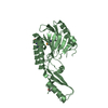
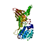
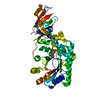
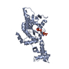
CRYSTAL STRUCTURE OF MRE11 FROM THERMOTOGA MARITIMA MSB8 (TM1635) AT 2.20 A RESOLUTION
ComponentsExonuclease, putative
KeywordsHYDROLASE / STRUCTURAL GENOMICS / JOINT CENTER FOR STRUCTURAL GENOMICS / JCSG / PROTEIN STRUCTURE INITIATIVE / PSI-2
Function / homology
Function and homology information


DNA exonuclease activity / 3'-5' exonuclease activity / DNA endonuclease activity / double-strand break repair / DNA recombination / DNA replication / Hydrolases; Acting on ester bonds / DNA repair / DNA binding / metal ion binding
Similarity search - Function
DNA double-strand break repair nuclease / Nuclease SbcCD subunit D / Mre11 nuclease, N-terminal metallophosphatase domain / Metallo-dependent phosphatases / Purple Acid Phosphatase; chain A, domain 2 / Calcineurin-like phosphoesterase domain, ApaH type / Calcineurin-like phosphoesterase / Metallo-dependent phosphatase-like / Double Stranded RNA Binding Domain / 4-Layer Sandwich ...DNA double-strand break repair nuclease / Nuclease SbcCD subunit D / Mre11 nuclease, N-terminal metallophosphatase domain / Metallo-dependent phosphatases / Purple Acid Phosphatase; chain A, domain 2 / Calcineurin-like phosphoesterase domain, ApaH type / Calcineurin-like phosphoesterase / Metallo-dependent phosphatase-like / Double Stranded RNA Binding Domain / 4-Layer Sandwich / 2-Layer Sandwich / Alpha Beta
Similarity search - Domain/homology
DNA double-strand break repair protein Mre11
Similarity search - Component
Biological speciesThermotoga maritima MSB8 (bacteria)
MethodX-RAY DIFFRACTION / SYNCHROTRON / MAD / Resolution: 2.2 Å
AuthorsJoint Center for Structural Genomics (JCSG)
CitationJournal: J.Mol.Biol. / Year: 2010
Title: Crystal structure of the first eubacterial Mre11 nuclease reveals novel features that may discriminate substrates during DNA repair.
Authors: Das, D. / Moiani, D. / Axelrod, H.L. / Miller, M.D. / McMullan, D. / Jin, K.K. / Abdubek, P. / Astakhova, T. / Burra, P. / Carlton, D. / Chiu, H.J. / Clayton, T. / Deller, M.C. / Duan, L. / ...Authors: Das, D. / Moiani, D. / Axelrod, H.L. / Miller, M.D. / McMullan, D. / Jin, K.K. / Abdubek, P. / Astakhova, T. / Burra, P. / Carlton, D. / Chiu, H.J. / Clayton, T. / Deller, M.C. / Duan, L. / Ernst, D. / Feuerhelm, J. / Grant, J.C. / Grzechnik, A. / Grzechnik, S.K. / Han, G.W. / Jaroszewski, L. / Klock, H.E. / Knuth, M.W. / Kozbial, P. / Krishna, S.S. / Kumar, A. / Marciano, D. / Morse, A.T. / Nigoghossian, E. / Okach, L. / Paulsen, J. / Reyes, R. / Rife, C.L. / Sefcovic, N. / Tien, H.J. / Trame, C.B. / van den Bedem, H. / Weekes, D. / Xu, Q. / Hodgson, K.O. / Wooley, J. / Elsliger, M.A. / Deacon, A.M. / Godzik, A. / Lesley, S.A. / Tainer, J.A. / Wilson, I.A.
History
DepositionJun 11, 2007Deposition site: RCSB / Processing site: RCSB
Revision 1.0Jun 26, 2007Provider: repository / Type: Initial release
Revision 1.1May 1, 2008Group: Version format compliance
Revision 1.2Jul 13, 2011Group: Advisory / Derived calculations / Version format compliance
Revision 1.3Oct 18, 2017Group: Refinement description / Category: software / Item: _software.classification / _software.name
Revision 1.4Oct 25, 2017Group: Author supporting evidence / Category: pdbx_struct_assembly_auth_evidence
Revision 1.5Jul 24, 2019Group: Data collection / Derived calculations / Refinement description
Category: software / struct_conn
Item: _software.classification / _software.contact_author ..._software.classification / _software.contact_author / _software.contact_author_email / _software.language / _software.location / _software.name / _software.type / _software.version / _struct_conn.pdbx_leaving_atom_flag
Revision 1.6Jan 25, 2023Group: Database references / Category: database_2 / struct_ref_seq_dif
Item: _database_2.pdbx_DOI / _database_2.pdbx_database_accession / _struct_ref_seq_dif.details
Remark 300 BIOMOLECULE: 1 THIS ENTRY CONTAINS THE CRYSTALLOGRAPHIC ASYMMETRIC UNIT WHICH CONSISTS OF 2 CHAINS. ... BIOMOLECULE: 1 THIS ENTRY CONTAINS THE CRYSTALLOGRAPHIC ASYMMETRIC UNIT WHICH CONSISTS OF 2 CHAINS. SEE REMARK 350 FOR INFORMATION ON GENERATING THE BIOLOGICAL MOLECULE(S). SIZE EXCLUSION CHROMATOGRAPHY SUGGESTS THE ASSIGNMENT OF A MONOMER AS THE SIGNIFICANT OLIGOMERIZATION STATE.
Remark 999 SEQUENCE THIS CONSTRUCT IS COMPRISED OF AMINO ACIDS 1-324 OF THE FULL-LENGTH PROTEIN (1-385) AND ... SEQUENCE THIS CONSTRUCT IS COMPRISED OF AMINO ACIDS 1-324 OF THE FULL-LENGTH PROTEIN (1-385) AND WAS EXPRESSED WITH A PURIFICATION TAG MGSDKIHHHHHH. DNA SEQUENCING INDICATES THAT RESIDUE 1 IS VALINE (NOT MET) IN THE CLONED CONSTRUCT. THE SEQUENCING RESULTS ARE CONSISTENT WITH THE ELECTRON DENSITY AND MASS SPECTROMETRY RESULTS FOR THE EXPRESSED PROTEIN.

-
Structure visualization

Structure viewerMolecule:
MolmilJmol/JSmol

Downloads & links

-
Assembly

Deposited unit
A: Exonuclease, putative
B: Exonuclease, putative


Theoretical massNumber of molelcules
Total (without water)77,7132
Polymers77,7132
Non-polymers00
Water2,864159
1
A: Exonuclease, putative


Theoretical massNumber of molelcules
Total (without water)38,8571
Polymers38,8571
Non-polymers00
Water181
TypeNameSymmetry operationNumber
identity operation1_555x,y,z1
MethodPQS
2
B: Exonuclease, putative


Theoretical massNumber of molelcules
Total (without water)38,8571
Polymers38,8571
Non-polymers00
Water181
TypeNameSymmetry operationNumber
identity operation1_555x,y,z1
MethodPQS
3


  • Idetical with deposited unit
  • defined by software
TypeNameSymmetry operationNumber
identity operation1_555x,y,z1
Buried area1580 Å2
ΔGint-23 kcal/mol
Surface area28020 Å2
MethodPISA
Unit cell
Length a, b, c (Å)47.598, 113.414, 80.951
Angle α, β, γ (deg.)90.000, 101.770, 90.000
Int Tables number4
Space group name H-MP1211
Noncrystallographic symmetry (NCS)NCS domain:
IDEns-IDDetails
11A
21B
31A
41B
51A
61B
71A
81B
91A
101B
111A
121B
131A
141B
151A
161B
171A
181B
191A
201B

NCS domain segments:

Ens-ID: 1 / Refine code: 4

Dom-IDComponent-IDBeg label comp-IDEnd label comp-IDAuth asym-IDLabel asym-IDAuth seq-IDLabel seq-ID
11LEULEUAA4 - 716 - 19
21LEULEUBB719
32LYSILEAA8 - 920 - 21
42LYSILEBB8 - 920 - 21
53LEUSERAA10 - 11022 - 122
63LEUSERBB10 - 11022 - 122
74SERASPAA111 - 112123 - 124
84SERASPBB111 - 112123 - 124
95ILESERAA113 - 118125 - 130
105ILESERBB113 - 118125 - 130
116PHEGLUAA119 - 120131 - 132
126PHEGLUBB119 - 120131 - 132
137PROASPAA121 - 245133 - 257
147PROASPBB121 - 245133 - 257
158GLULYSAA246 - 247258 - 259
168GLULYSBB246 - 247258 - 259
179GLYGLUAA248 - 263260 - 275
189GLYGLUBB248 - 263260 - 275
1910ARGGLUAA264 - 324276 - 336
2010ARGGLUBB264 - 324276 - 336
DetailsSIZE EXCLUSION CHROMATOGRAPHY SUGGESTS THE ASSIGNMENT OF A MONOMER AS THE SIGNIFICANT OLIGOMERIZATION STATE.

-
Components

#1: Protein Exonuclease, putative /


Mass: 38856.582 Da / Num. of mol.: 2 / Fragment: Residues 1-324
Source method: isolated from a genetically manipulated source
Source: (gene. exp.) Thermotoga maritima MSB8 (bacteria) / Species: Thermotoga maritima / Strain: MSB8, DSM 3109, JCM 10099 / Gene: TM_1635 / Plasmid: speedET / Production host: Escherichia coli (E. coli) / Strain (production host): HK100 / References: UniProt: Q9X1X0
#2: Water ChemComp-HOH / water / Water


Mass: 18.015 Da / Num. of mol.: 159 / Source method: isolated from a natural source / Formula: H2O

-
Experimental details

-
Experiment

ExperimentMethod: X-RAY DIFFRACTION / Number of used crystals: 2

-
Sample preparation

Crystal
IDDensity Matthews3/Da)Density % sol (%)Description
12.7555.29TWO CRYSTALS WERE USED FOR THE SOLUTION OF THIS STRUCTURE. A 2.4 ANGSTROM MAD DATA COLLECTED FROM ONE CRYSTAL WAS USED TO PHASE AND TRACE AN INITIAL MODEL. THE MODEL WAS THEN REFINED USING THE AMPLITUDES FROM A SECOND CRYSTAL THAT DIFFRACTED TO AN ENHANCED RESOLUTION OF 2.2 ANGSTROM. THE 2.4 ANGSTROM MAD PHASES FROM THE FIRST CRYSTAL WERE USED AS PHASE RESTRAINTS DURING THE REFINEMENT.
2
Crystal grow
Temperature (K)Crystal-IDMethodpHDetails
2771vapor diffusion, sitting drop5.57NANODROP, 10.3% PEG 6000, 0.1M MES pH 5.57, VAPOR DIFFUSION, SITTING DROP, temperature 277K
2772vapor diffusion, sitting drop5.14NANODROP, 2.5% PEG 6000, 0.1M MES pH 5.14, VAPOR DIFFUSION, SITTING DROP, temperature 277K

-
Data collection

Diffraction
IDMean temperature (K)Crystal-ID
11001
22
Diffraction source
SourceSiteBeamlineIDWavelength (Å)
SYNCHROTRONSSRL BL11-110.98086
SYNCHROTRONSSRL BL11-120.91837, 0.97917, 0.97891
DetectorType: MARMOSAIC 325 mm CCD / Detector: CCD / Date: May 13, 2007 / Details: Flat mirror (vertical focusing)
RadiationMonochromator: Single crystal Si(111) bent (horizontal focusing)
Protocol: MAD / Monochromatic (M) / Laue (L): M / Scattering type: x-ray
Radiation wavelength
IDWavelength (Å)Relative weight
10.980861
20.918371
30.979171
40.978911
ReflectionResolution: 2.2→37.45 Å / Num. obs: 56132 / % possible obs: 98.8 % / Redundancy: 2.6 % / Biso Wilson estimate: 43.03 Å2 / Rmerge(I) obs: 0.043 / Rsym value: 0.043 / Net I/σ(I): 11.3
Reflection shell

Diffraction-ID: 1

Resolution (Å)Redundancy (%)Rmerge(I) obsMean I/σ(I) obsNum. measured allNum. unique allRsym value% possible all
2.2-2.262.50.5330.9798331320.53399.5
2.26-2.322.60.4241.7790730700.42499.4
2.32-2.392.60.3532.1758429660.35399.7
2.39-2.462.60.2862.5740028680.28699.4
2.46-2.542.60.2313.1724828040.23199.5
2.54-2.632.60.184692926950.1899.5
2.63-2.732.60.1514.6669225940.15199.4
2.73-2.842.60.1166.2654825390.11699.4
2.84-2.972.60.0858.2622924130.08599.4
2.97-3.112.60.06810.3596922910.06899.4
3.11-3.282.60.05312.3564921820.05399.4
3.28-3.482.60.04315537320740.04399.1
3.48-3.722.60.03714.8507019550.03799.1
3.72-4.022.60.03317.1472618050.03398.6
4.02-4.42.60.02621.9431816430.02698.2
4.4-4.922.60.02221.9392214890.02297.5
4.92-5.682.70.02317.8350613110.02396.3
5.68-6.962.70.02225.1290110840.02295.4
6.96-9.842.70.02224.222248220.02292.6
9.84-37.452.60.02326.211134230.02384.7

-
Phasing

PhasingMethod: MAD

-
Processing

Software
NameVersionClassificationNB
REFMAC5.2.0019refinement
PHENIXrefinement
SHELXphasing
MolProbity3beta29model building
SCALAdata scaling
PDB_EXTRACT3data extraction
MAR345CCDdata collection
MOSFLMdata reduction
SHELXDphasing
autoSHARPphasing
RefinementMethod to determine structure: MAD / Resolution: 2.2→37.45 Å / Cor.coef. Fo:Fc: 0.959 / Cor.coef. Fo:Fc free: 0.939 / SU B: 12.776 / SU ML: 0.162 / TLS residual ADP flag: LIKELY RESIDUAL / Cross valid method: THROUGHOUT / σ(F): 0 / ESU R: 0.223 / ESU R Free: 0.191
Stereochemistry target values: MAXIMUM LIKELIHOOD WITH PHASES
Details: 1. HYDROGENS HAVE BEEN ADDED IN THE RIDING POSITIONS. 2. A MET-INHIBITION PROTOCOL WAS USED FOR SELENOMETHIONINE INCORPORATION DURING PROTEIN EXPRESSION. THE OCCUPANCY OF THE SE ATOMS IN THE ...Details: 1. HYDROGENS HAVE BEEN ADDED IN THE RIDING POSITIONS. 2. A MET-INHIBITION PROTOCOL WAS USED FOR SELENOMETHIONINE INCORPORATION DURING PROTEIN EXPRESSION. THE OCCUPANCY OF THE SE ATOMS IN THE MSE RESIDUES WAS REDUCED TO 0.75 TO ACCOUNT FOR THE REDUCED SCATTERING POWER DUE TO PARTIAL S-MET INCORPORATION. 3. ATOM RECORD CONTAINS RESIDUAL B FACTORS ONLY. 4. RESIDUES A93-A98, A141-A147, A188-A195, B95-B98, B141-B149, B187-B195, B311-B312 ARE DISORDERED AND ARE NOT MODELED. 5. THE STRUCTURE FACTOR AMPLITUDES USED IN THE FINAL REFINEMENT WERE FROM A NATIVE CRYSTAL. THE REFINEMENT OF THE COORDINATES WAS RESTRAINED WITH THE EXPERIMENTAL PHASES FROM A CRYSTAL OF THE SELENOMETHIONINE-SUBSTITUTED PROTEIN THAT WAS USED FOR INITIAL PHASE DETERMINATION BY MULTIPLE WAVELENGTH ANOMALOUS DISPERSION.
RfactorNum. reflection% reflectionSelection details
Rfree0.241 2120 5 %RANDOM
Rwork0.198 ---
obs0.2 42148 98.71 %-
Solvent computationIon probe radii: 0.8 Å / Shrinkage radii: 0.8 Å / VDW probe radii: 1.2 Å / Solvent model: MASK
Displacement parametersBiso mean: 41.949 Å2
Baniso -1Baniso -2Baniso -3
1--2.12 Å20 Å20.5 Å2
2--2.73 Å20 Å2
3----0.41 Å2
Refinement stepCycle: LAST / Resolution: 2.2→37.45 Å
ProteinNucleic acidLigandSolventTotal
Num. atoms4778 0 0 159 4937
Refine LS restraints
Refine-IDTypeDev idealDev ideal targetNumber
X-RAY DIFFRACTIONr_bond_refined_d0.0160.0224978
X-RAY DIFFRACTIONr_bond_other_d0.0020.023452
X-RAY DIFFRACTIONr_angle_refined_deg1.4281.9776756
X-RAY DIFFRACTIONr_angle_other_deg0.81738417
X-RAY DIFFRACTIONr_dihedral_angle_1_deg6.6735620
X-RAY DIFFRACTIONr_dihedral_angle_2_deg34.06923.423222
X-RAY DIFFRACTIONr_dihedral_angle_3_deg15.89815877
X-RAY DIFFRACTIONr_dihedral_angle_4_deg19.6271534
X-RAY DIFFRACTIONr_chiral_restr0.0650.2769
X-RAY DIFFRACTIONr_gen_planes_refined0.0150.025450
X-RAY DIFFRACTIONr_gen_planes_other0.0020.021024
X-RAY DIFFRACTIONr_nbd_refined0.2330.3896
X-RAY DIFFRACTIONr_nbd_other0.2180.33526
X-RAY DIFFRACTIONr_nbtor_refined0.1910.52373
X-RAY DIFFRACTIONr_nbtor_other0.0930.52724
X-RAY DIFFRACTIONr_xyhbond_nbd_refined0.2140.5350
X-RAY DIFFRACTIONr_xyhbond_nbd_other0.1350.53
X-RAY DIFFRACTIONr_symmetry_vdw_refined0.0760.312
X-RAY DIFFRACTIONr_symmetry_vdw_other0.3150.329
X-RAY DIFFRACTIONr_symmetry_hbond_refined0.1920.58
X-RAY DIFFRACTIONr_mcbond_it4.03833120
X-RAY DIFFRACTIONr_mcbond_other1.47731215
X-RAY DIFFRACTIONr_mcangle_it5.28354950
X-RAY DIFFRACTIONr_scbond_it8.36182073
X-RAY DIFFRACTIONr_scangle_it10.128111792
Refine LS restraints NCS

Dom-ID: 1 / Auth asym-ID: A / Ens-ID: 1 / Number: 3827 / Refine-ID: X-RAY DIFFRACTION

TypeRms dev position (Å)Weight position
MEDIUM POSITIONAL0.550.5
MEDIUM THERMAL2.22
LS refinement shellResolution: 2.2→2.257 Å / Total num. of bins used: 20
RfactorNum. reflection% reflection
Rfree0.371 156 -
Rwork0.272 2970 -
obs-3126 99.3 %
Refinement TLS params.

Method: refined / Refine-ID: X-RAY DIFFRACTION

IDL112)L122)L132)L222)L232)L332)S11 (Å °)S12 (Å °)S13 (Å °)S21 (Å °)S22 (Å °)S23 (Å °)S31 (Å °)S32 (Å °)S33 (Å °)T112)T122)T132)T222)T232)T332)Origin x (Å)Origin y (Å)Origin z (Å)
11.3086-0.53820.25462.129-0.89041.09920.0637-0.02430.124-0.1556-0.1071-0.01020.04570.08230.0434-0.1836-0.03190.0111-0.0852-0.0764-0.1126-17.3458-96.0634-0.9736
22.39360.48710.75691.72942.24215.53880.06650.14950.00550.0966-0.17780.08970.2534-0.6720.1113-0.1081-0.03810.012-0.07230.0449-0.0711-32.3899-100.468941.2857
Refinement TLS group
IDRefine-IDRefine TLS-IDAuth asym-IDLabel asym-IDAuth seq-IDLabel seq-ID
1X-RAY DIFFRACTION1AA4 - 32416 - 336
2X-RAY DIFFRACTION2BB7 - 32419 - 336

+
About Yorodumi

-
News

-
Feb 9, 2022. New format data for meta-information of EMDB entries

New format data for meta-information of EMDB entries

  • Version 3 of the EMDB header file is now the official format.
  • The previous official version 1.9 will be removed from the archive.

Related info.:EMDB header

External links:wwPDB to switch to version 3 of the EMDB data model

-
Aug 12, 2020. Covid-19 info

Covid-19 info

URL: https://pdbj.org/emnavi/covid19.php

New page: Covid-19 featured information page in EM Navigator.

Related info.:Covid-19 info / Mar 5, 2020. Novel coronavirus structure data

+
Mar 5, 2020. Novel coronavirus structure data

Novel coronavirus structure data

Related info.:Yorodumi Speices / Aug 12, 2020. Covid-19 info

External links:COVID-19 featured content - PDBj / Molecule of the Month (242):Coronavirus Proteases

+
Jan 31, 2019. EMDB accession codes are about to change! (news from PDBe EMDB page)

EMDB accession codes are about to change! (news from PDBe EMDB page)

  • The allocation of 4 digits for EMDB accession codes will soon come to an end. Whilst these codes will remain in use, new EMDB accession codes will include an additional digit and will expand incrementally as the available range of codes is exhausted. The current 4-digit format prefixed with “EMD-” (i.e. EMD-XXXX) will advance to a 5-digit format (i.e. EMD-XXXXX), and so on. It is currently estimated that the 4-digit codes will be depleted around Spring 2019, at which point the 5-digit format will come into force.
  • The EM Navigator/Yorodumi systems omit the EMD- prefix.

Related info.:Q: What is EMD? / ID/Accession-code notation in Yorodumi/EM Navigator

External links:EMDB Accession Codes are Changing Soon! / Contact to PDBj

+
Jul 12, 2017. Major update of PDB

Major update of PDB

  • wwPDB released updated PDB data conforming to the new PDBx/mmCIF dictionary.
  • This is a major update changing the version number from 4 to 5, and with Remediation, in which all the entries are updated.
  • In this update, many items about electron microscopy experimental information are reorganized (e.g. em_software).
  • Now, EM Navigator and Yorodumi are based on the updated data.

External links:wwPDB Remediation / Enriched Model Files Conforming to OneDep Data Standards Now Available in the PDB FTP Archive

-
Yorodumi

Thousand views of thousand structures

  • Yorodumi is a browser for structure data from EMDB, PDB, SASBDB, etc.
  • This page is also the successor to EM Navigator detail page, and also detail information page/front-end page for Omokage search.
  • The word "yorodu" (or yorozu) is an old Japanese word meaning "ten thousand". "mi" (miru) is to see.

Related info.:EMDB / PDB / SASBDB / Comparison of 3 databanks / Yorodumi Search / Aug 31, 2016. New EM Navigator & Yorodumi / Yorodumi Papers / Jmol/JSmol / Function and homology information / Changes in new EM Navigator and Yorodumi

Read more