 Movie
Movie Controller

[English] 日本語
 Yorodumi
- PDB-6ap0: Crystal structure of human FLASH N-terminal domain C54S/C83A (Cry... -
+ Open data

- Basic information
| Entry | Database: PDB / ID: 6ap0 | ||||||
|---|---|---|---|---|---|---|---|
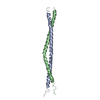
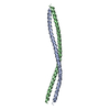
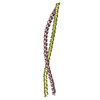
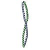
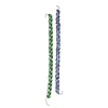
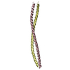
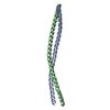
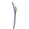
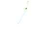
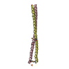
| Title | Crystal structure of human FLASH N-terminal domain C54S/C83A (Crystal form 2) | ||||||
 Components Components | CASP8-associated protein 2 | ||||||
 Keywords Keywords |  GENE REGULATION / coiled-coil GENE REGULATION / coiled-coil | ||||||
| Function / homology |  Function and homology information Function and homology informationFas signaling pathway / SUMO polymer binding / peptidase activator activity involved in apoptotic process / death receptor binding / cysteine-type endopeptidase activator activity involved in apoptotic process / extrinsic apoptotic signaling pathway via death domain receptors / SUMOylation of transcription cofactors / apoptotic signaling pathway / PML body / cellular response to mechanical stimulus ...Fas signaling pathway / SUMO polymer binding / peptidase activator activity involved in apoptotic process / death receptor binding / cysteine-type endopeptidase activator activity involved in apoptotic process / extrinsic apoptotic signaling pathway via death domain receptors / SUMOylation of transcription cofactors / apoptotic signaling pathway / PML body / cellular response to mechanical stimulus / transcription corepressor activity / activation of cysteine-type endopeptidase activity involved in apoptotic process / cell cycle / signal transduction / mitochondrion / nucleoplasm / nucleus / cytoplasmSimilarity search - Function | ||||||
| Biological species |  Homo sapiens (human) Homo sapiens (human) | ||||||
| Method | X-RAY DIFFRACTION / SYNCHROTRON / MOLECULAR REPLACEMENT / Resolution: 2.581 Å | ||||||
 Authors Authors | Aik, W.S. / Tong, L. | ||||||
 Citation Citation | Journal: PLoS ONE / Year: 2017 Title: The N-terminal domains of FLASH and Lsm11 form a 2:1 heterotrimer for histone pre-mRNA 3'-end processing. Authors: Aik, W.S. / Lin, M.H. / Tan, D. / Tripathy, A. / Marzluff, W.F. / Dominski, Z. / Chou, C.Y. / Tong, L. | ||||||
| History |
|
- Structure visualization
| Structure viewer | Molecule: MolmilJmol/JSmol |
|---|
- Downloads & links
-Download
| PDBx/mmCIF format | 6ap0.cif.gz | 84.1 KB | Display | PDBx/mmCIF format |
|---|---|---|---|---|
| PDB format | pdb6ap0.ent.gz | 64.2 KB | Display | PDB format |
| PDBx/mmJSON format | 6ap0.json.gz | Tree view | PDBx/mmJSON format | |
| Others |  Other downloads Other downloads |
-Validation report
| Arichive directory | https://data.pdbj.org/pub/pdb/validation_reports/ap/6ap0ftp://data.pdbj.org/pub/pdb/validation_reports/ap/6ap0 | HTTPS FTP |
|---|
-Related structure data
| Related structure data |  6anoSC 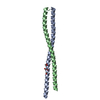 6aozC S: Starting model for refinement C: citing same article ( |
|---|---|
| Similar structure data |
-Links
 PDBj
PDBj- Assembly
| Deposited unit | 
| ||||||||
|---|---|---|---|---|---|---|---|---|---|
| 1 |
| ||||||||
| Unit cell |
|
-Components
| #1: Protein | Mass: 11333.769 Da / Num. of mol.: 2 / Fragment: UNP residues 51-137 / Mutation: C54S, C83A Source method: isolated from a genetically manipulated source Source: (gene. exp.) Homo sapiens (human) / Gene: CASP8AP2, FLASH, KIAA1315, RIP25 / Production host:  Escherichia coli (E. coli) / References: UniProt: Q9UKL3 Escherichia coli (E. coli) / References: UniProt: Q9UKL3#2: Water | ChemComp-HOH / | Water Mass: 18.015 Da / Num. of mol.: 10 / Source method: isolated from a natural source / Formula: H2O Mass: 18.015 Da / Num. of mol.: 10 / Source method: isolated from a natural source / Formula: H2O |
|---|
-Experimental details
-Experiment
| Experiment | Method: X-RAY DIFFRACTION / Number of used crystals: 1 |
|---|
- Sample preparation
| Crystal | Density Matthews: 2.1 Å3/Da / Density % sol: 41.44 % |
|---|---|
| Crystal grow | Temperature: 293 K / Method: vapor diffusion, sitting drop Details: 100 mM sodium formate, 15% (w/v) PEG 3350, 3% (v/v) 1,6-hexanediol |
-Data collection
| Diffraction | Mean temperature: 100 K |
|---|---|
| Diffraction source | Source: SYNCHROTRON / Site: APS  / Beamline: 24-ID-E / Wavelength: 0.9792 Å / Beamline: 24-ID-E / Wavelength: 0.9792 Å |
| Detector | Type: ADSC QUANTUM 315 / Detector: CCD / Date: Mar 10, 2016 |
| Radiation | Protocol: SINGLE WAVELENGTH / Monochromatic (M) / Laue (L): M / Scattering type: x-ray |
| Radiation wavelength | Wavelength: 0.9792 Å / Relative weight: 1 |
| Reflection | Resolution: 2.58→36.216 Å / Num. obs: 11542 / % possible obs: 99.6 % / Redundancy: 3.8 % / CC1/2: 0.998 / Rmerge(I) obs: 0.079 / Net I/σ(I): 13.42 |
| Reflection shell | Resolution: 2.58→2.74 Å / Redundancy: 3.7 % / Rmerge(I) obs: 0.467 / Mean I/σ(I) obs: 3.82 / Num. unique obs: 1848 / CC1/2: 0.898 / % possible all: 99.2 |
- Processing
| Software |
| ||||||||||||||||||||||||||||||||||||||||||||||||||||||||||||||||||||||||||||||||||||||||||||||||||||
|---|---|---|---|---|---|---|---|---|---|---|---|---|---|---|---|---|---|---|---|---|---|---|---|---|---|---|---|---|---|---|---|---|---|---|---|---|---|---|---|---|---|---|---|---|---|---|---|---|---|---|---|---|---|---|---|---|---|---|---|---|---|---|---|---|---|---|---|---|---|---|---|---|---|---|---|---|---|---|---|---|---|---|---|---|---|---|---|---|---|---|---|---|---|---|---|---|---|---|---|---|---|
| Refinement | Method to determine structure: MOLECULAR REPLACEMENT Starting model: 6ANO Resolution: 2.581→36.216 Å / SU ML: 0.37 / Cross valid method: FREE R-VALUE / σ(F): 1.35 / Phase error: 27
| ||||||||||||||||||||||||||||||||||||||||||||||||||||||||||||||||||||||||||||||||||||||||||||||||||||
| Solvent computation | Shrinkage radii: 0.9 Å / VDW probe radii: 1.11 Å | ||||||||||||||||||||||||||||||||||||||||||||||||||||||||||||||||||||||||||||||||||||||||||||||||||||
| Refinement step | Cycle: LAST / Resolution: 2.581→36.216 Å
| ||||||||||||||||||||||||||||||||||||||||||||||||||||||||||||||||||||||||||||||||||||||||||||||||||||
| Refine LS restraints |
| ||||||||||||||||||||||||||||||||||||||||||||||||||||||||||||||||||||||||||||||||||||||||||||||||||||
| LS refinement shell |
| ||||||||||||||||||||||||||||||||||||||||||||||||||||||||||||||||||||||||||||||||||||||||||||||||||||
| Refinement TLS params. | Method: refined / Refine-ID: X-RAY DIFFRACTION
| ||||||||||||||||||||||||||||||||||||||||||||||||||||||||||||||||||||||||||||||||||||||||||||||||||||
| Refinement TLS group |
|
