 Movie
Movie Controller


+ Open data

- Basic information
| Entry | Database: PDB / ID: 5k7b | |||||||||||||||
|---|---|---|---|---|---|---|---|---|---|---|---|---|---|---|---|---|
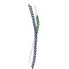
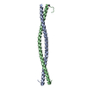
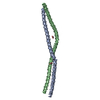
| Title | Beclin 2 CCD homodimer | |||||||||||||||
 Components Components | Beclin-2 | |||||||||||||||
 Keywords Keywords |  APOPTOSIS / Coiled-coil Domain / Autophagy APOPTOSIS / Coiled-coil Domain / Autophagy | |||||||||||||||
| Function / homology |  Function and homology information Function and homology informationG protein-coupled receptor catabolic process / phosphatidylinositol 3-kinase complex, class III, type II / phosphatidylinositol 3-kinase complex, class III, type I / cellular response to nitrogen starvation / late endosome to vacuole transport / phagophore assembly site / endosome to lysosome transport / autophagosome assembly / autophagySimilarity search - Function | |||||||||||||||
| Biological species |  Homo sapiens (human) Homo sapiens (human) | |||||||||||||||
| Method | X-RAY DIFFRACTION / SYNCHROTRON / MOLECULAR REPLACEMENT / Resolution: 2.3 Å | |||||||||||||||
 Authors Authors | Su, M. / Sinha, S. | |||||||||||||||
| Funding support |  United States, 4items United States, 4items
| |||||||||||||||
 Citation Citation | Journal: Protein Sci. / Year: 2017 Title: BECN2 interacts with ATG14 through a metastable coiled-coil to mediate autophagy. Authors: Su, M. / Li, Y. / Wyborny, S. / Neau, D. / Chakravarthy, S. / Levine, B. / Colbert, C.L. / Sinha, S.C. | |||||||||||||||
| History |
|
- Structure visualization
| Structure viewer | Molecule: MolmilJmol/JSmol |
|---|
- Downloads & links
-Download
| PDBx/mmCIF format | 5k7b.cif.gz | 153.8 KB | Display | PDBx/mmCIF format |
|---|---|---|---|---|
| PDB format | pdb5k7b.ent.gz | 123.5 KB | Display | PDB format |
| PDBx/mmJSON format | 5k7b.json.gz | Tree view | PDBx/mmJSON format | |
| Others |  Other downloads Other downloads |
-Validation report
| Arichive directory | https://data.pdbj.org/pub/pdb/validation_reports/k7/5k7bftp://data.pdbj.org/pub/pdb/validation_reports/k7/5k7b | HTTPS FTP |
|---|
-Related structure data


-Links
 PDBj
PDBj
- Assembly
| Deposited unit | 
| ||||||||
|---|---|---|---|---|---|---|---|---|---|
| 1 | 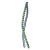
| ||||||||
| 2 | 
| ||||||||
| Unit cell |
|
-Components
| #1: Protein | Mass: 11300.361 Da / Num. of mol.: 4 / Fragment: UNP residues 158-250 Source method: isolated from a genetically manipulated source Source: (gene. exp.) Homo sapiens (human) / Gene: BECN2, BECN1L1, BECN1P1 / Production host:  Escherichia coli (E. coli) / References: UniProt: A8MW95 Escherichia coli (E. coli) / References: UniProt: A8MW95#2: Water | ChemComp-HOH / | Water Mass: 18.015 Da / Num. of mol.: 139 / Source method: isolated from a natural source / Formula: H2O Mass: 18.015 Da / Num. of mol.: 139 / Source method: isolated from a natural source / Formula: H2O |
|---|
-Experimental details
-Experiment
| Experiment | Method: X-RAY DIFFRACTION / Number of used crystals: 1 |
|---|
- Sample preparation
| Crystal | Density Matthews: 2.28 Å3/Da / Density % sol: 45.96 % |
|---|---|
| Crystal grow | Temperature: 293.15 K / Method: vapor diffusion, hanging drop / pH: 6.5 Details: 0.1 M Bis-Tris pH 6.5, 0.1 M NaCl and 1.5 M (NH4)2SO4 |
-Data collection
| Diffraction | Mean temperature: 100 K |
|---|---|
| Diffraction source | Source: SYNCHROTRON / Site: APS / Beamline: 24-ID-E / Wavelength: 0.9792 Å |
| Detector | Type: ADSC QUANTUM 315 / Detector: CCD / Date: Nov 5, 2012 |
| Radiation | Protocol: SINGLE WAVELENGTH / Monochromatic (M) / Laue (L): M / Scattering type: x-ray |
| Radiation wavelength | Wavelength: 0.9792 Å / Relative weight: 1 |
| Reflection | Resolution: 2.19→40.33 Å / Num. obs: 17947 / % possible obs: 84.6 % / Redundancy: 3.4 % / CC1/2: 0.997 / Net I/σ(I): 7.7 |
| Reflection shell | Resolution: 2.19→2.26 Å / Redundancy: 3.3 % / Mean I/σ(I) obs: 2.6 / % possible all: 87 |
- Processing
| Software |
| |||||||||||||||||||||||||||||||||||||||||||||||||
|---|---|---|---|---|---|---|---|---|---|---|---|---|---|---|---|---|---|---|---|---|---|---|---|---|---|---|---|---|---|---|---|---|---|---|---|---|---|---|---|---|---|---|---|---|---|---|---|---|---|---|
| Refinement | Method to determine structure: MOLECULAR REPLACEMENT / Resolution: 2.3→40.33 Å / Cross valid method: FREE R-VALUE
| |||||||||||||||||||||||||||||||||||||||||||||||||
| Refinement step | Cycle: LAST / Resolution: 2.3→40.33 Å
| |||||||||||||||||||||||||||||||||||||||||||||||||
| Refine LS restraints |
| |||||||||||||||||||||||||||||||||||||||||||||||||
| LS refinement shell |
| |||||||||||||||||||||||||||||||||||||||||||||||||
| Refinement TLS params. | Method: refined / Origin x: 44.981 Å / Origin y: 28.763 Å / Origin z: 10.585 Å
| |||||||||||||||||||||||||||||||||||||||||||||||||
| Refinement TLS group |
|
