 Movie
Movie Controller


+ Open data

- Basic information
| Entry | Database: PDB / ID: 6a4v | |||||||||
|---|---|---|---|---|---|---|---|---|---|---|
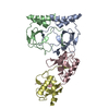
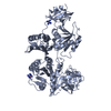
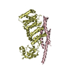
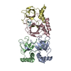
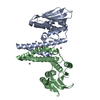
| Title | Open Reading frame 49 | |||||||||
 Components Components | 49 protein | |||||||||
 Keywords Keywords |  REPLICATION / Gamma herpesvirus 68 Open reading frame 49 / ORF49 Poly (ADP-ribose) polymerase-1 (PARP-1) REPLICATION / Gamma herpesvirus 68 Open reading frame 49 / ORF49 Poly (ADP-ribose) polymerase-1 (PARP-1) | |||||||||
| Function / homology | BRRF1, herpesviridae / BRRF1-like protein / 49 protein Function and homology information Function and homology information | |||||||||
| Biological species |  Murid herpesvirus 4 (Murine herpesvirus 68) Murid herpesvirus 4 (Murine herpesvirus 68) | |||||||||
| Method | X-RAY DIFFRACTION / SYNCHROTRON / SAD / Resolution: 2.2 Å | |||||||||
 Authors Authors | Hwang, K.Y. / Song, M.J. / Kim, J.S. / Cheong, W.C. | |||||||||
 Citation Citation | Journal: Iucrj / Year: 2018 Title: Structure-based mechanism of action of a viral poly(ADP-ribose) polymerase 1-interacting protein facilitating virus replication. Authors: Chung, W.C. / Kim, J. / Kim, B.C. / Kang, H.R. / Son, J. / Ki, H. / Hwang, K.Y. / Song, M.J. | |||||||||
| History |
|
- Structure visualization
| Structure viewer | Molecule: MolmilJmol/JSmol |
|---|
- Downloads & links
-Download
| PDBx/mmCIF format | 6a4v.cif.gz | 123.5 KB | Display | PDBx/mmCIF format |
|---|---|---|---|---|
| PDB format | pdb6a4v.ent.gz | 96 KB | Display | PDB format |
| PDBx/mmJSON format | 6a4v.json.gz | Tree view | PDBx/mmJSON format | |
| Others |  Other downloads Other downloads |
-Validation report
| Arichive directory | https://data.pdbj.org/pub/pdb/validation_reports/a4/6a4vftp://data.pdbj.org/pub/pdb/validation_reports/a4/6a4v | HTTPS FTP |
|---|
-Related structure data
| Similar structure data |
|---|


-Links
 PDBj
PDBj- Assembly
| Deposited unit | 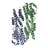
| ||||||||
|---|---|---|---|---|---|---|---|---|---|
| 1 |
| ||||||||
| Unit cell |
|
-Components
| #1: Protein | Mass: 38201.805 Da / Num. of mol.: 2 Source method: isolated from a genetically manipulated source Source: (gene. exp.) Murid herpesvirus 4 (Murine herpesvirus 68)Gene: BNLF1, 49, GAMMAHV.ORF49 / Production host:  Escherichia coli (E. coli) / References: UniProt: P88987 Escherichia coli (E. coli) / References: UniProt: P88987#2: Water | ChemComp-HOH / | Water Mass: 18.015 Da / Num. of mol.: 60 / Source method: isolated from a natural source / Formula: H2O Mass: 18.015 Da / Num. of mol.: 60 / Source method: isolated from a natural source / Formula: H2O |
|---|
-Experimental details
-Experiment
| Experiment | Method: X-RAY DIFFRACTION / Number of used crystals: 1 |
|---|
- Sample preparation
| Crystal | Density Matthews: 5.34 Å3/Da / Density % sol: 76.96 % |
|---|---|
| Crystal grow | Temperature: 293 K / Method: vapor diffusion, hanging drop / pH: 8.2 Details: 0.1M Tris-Hcl (pH 8.2), 0.33M sodium/potassium tartrate 0.5% polyehylene glycerol 5000 mono-methyl ether |
-Data collection
| Diffraction | Mean temperature: 100 K | |||||||||||||||
|---|---|---|---|---|---|---|---|---|---|---|---|---|---|---|---|---|
| Diffraction source | Source: SYNCHROTRON / Site: Photon Factory  / Beamline: BL-1A / Wavelength: 0.979 Å / Beamline: BL-1A / Wavelength: 0.979 Å | |||||||||||||||
| Detector | Type: DECTRIS PILATUS 2M / Detector: PIXEL / Date: Mar 5, 2012 | |||||||||||||||
| Radiation | Protocol: SINGLE WAVELENGTH / Monochromatic (M) / Laue (L): M / Scattering type: x-ray | |||||||||||||||
| Radiation wavelength | Wavelength: 0.979 Å / Relative weight: 1 | |||||||||||||||
| Reflection twin |
| |||||||||||||||
| Reflection | Resolution: 2.2→30 Å / Num. obs: 272254 / % possible obs: 90.5 % / Redundancy: 3.6 % / Biso Wilson estimate: 51.49 Å2 / Rmerge(I) obs: 0.079 / Net I/σ(I): 11.94 | |||||||||||||||
| Reflection shell | Resolution: 2.2→2.24 Å / Rmerge(I) obs: 0.414 / Num. unique obs: 7618 |
- Processing
| Software |
| ||||||||||||||||||||||||||||||||||||||||||||||||||||||||||||||||||||||||||||||||||||||||||||||||||||||||||||||||||||||||||||||||||||||||||||||||||||||||||||||||||||||||||||||||||||||
|---|---|---|---|---|---|---|---|---|---|---|---|---|---|---|---|---|---|---|---|---|---|---|---|---|---|---|---|---|---|---|---|---|---|---|---|---|---|---|---|---|---|---|---|---|---|---|---|---|---|---|---|---|---|---|---|---|---|---|---|---|---|---|---|---|---|---|---|---|---|---|---|---|---|---|---|---|---|---|---|---|---|---|---|---|---|---|---|---|---|---|---|---|---|---|---|---|---|---|---|---|---|---|---|---|---|---|---|---|---|---|---|---|---|---|---|---|---|---|---|---|---|---|---|---|---|---|---|---|---|---|---|---|---|---|---|---|---|---|---|---|---|---|---|---|---|---|---|---|---|---|---|---|---|---|---|---|---|---|---|---|---|---|---|---|---|---|---|---|---|---|---|---|---|---|---|---|---|---|---|---|---|---|---|
| Refinement | Method to determine structure: SAD / Resolution: 2.2→29.82 Å / Cor.coef. Fo:Fc: 0.932 / Cor.coef. Fo:Fc free: 0.91 / SU B: 2.682 / SU ML: 0.072 / Cross valid method: THROUGHOUT / ESU R: 0.031 / ESU R Free: 0.031 / Details: HYDROGENS HAVE BEEN ADDED IN THE RIDING POSITIONS
| ||||||||||||||||||||||||||||||||||||||||||||||||||||||||||||||||||||||||||||||||||||||||||||||||||||||||||||||||||||||||||||||||||||||||||||||||||||||||||||||||||||||||||||||||||||||
| Solvent computation | Ion probe radii: 0.8 Å / Shrinkage radii: 0.8 Å / VDW probe radii: 1.2 Å | ||||||||||||||||||||||||||||||||||||||||||||||||||||||||||||||||||||||||||||||||||||||||||||||||||||||||||||||||||||||||||||||||||||||||||||||||||||||||||||||||||||||||||||||||||||||
| Displacement parameters | Biso mean: 54.832 Å2
| ||||||||||||||||||||||||||||||||||||||||||||||||||||||||||||||||||||||||||||||||||||||||||||||||||||||||||||||||||||||||||||||||||||||||||||||||||||||||||||||||||||||||||||||||||||||
| Refinement step | Cycle: 1 / Resolution: 2.2→29.82 Å
| ||||||||||||||||||||||||||||||||||||||||||||||||||||||||||||||||||||||||||||||||||||||||||||||||||||||||||||||||||||||||||||||||||||||||||||||||||||||||||||||||||||||||||||||||||||||
| Refine LS restraints |
|