[English] 日本語
Yorodumi
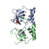
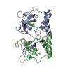
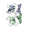
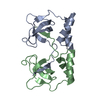
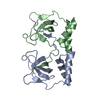
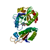
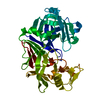
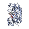
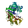
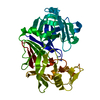
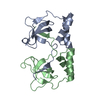
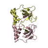
- PDB-2d7e: Crystal structure of N-terminal domain of PriA from E.coli -

+
Open data


ID or keywords:

Loading...

-
Basic information

Entry
Database: PDB / ID: 2d7e
TitleCrystal structure of N-terminal domain of PriA from E.coli
ComponentsPrimosomal protein N'
KeywordsHYDROLASE / inter-twined
Function / homology
Function and homology information


DnaB-DnaC-DnaT-PriA-PriC complex / DnaB-DnaC-DnaT-PriA-PriB complex / plasmid maintenance / primosome complex / DNA replication, synthesis of primer / 3'-5' DNA helicase activity / replication fork processing / DNA unwinding involved in DNA replication / DNA replication initiation / helicase activity ...DnaB-DnaC-DnaT-PriA-PriC complex / DnaB-DnaC-DnaT-PriA-PriB complex / plasmid maintenance / primosome complex / DNA replication, synthesis of primer / 3'-5' DNA helicase activity / replication fork processing / DNA unwinding involved in DNA replication / DNA replication initiation / helicase activity / response to gamma radiation / Hydrolases; Acting on acid anhydrides; Acting on acid anhydrides to facilitate cellular and subcellular movement / DNA-templated DNA replication / double-strand break repair / DNA recombination / DNA replication / hydrolase activity / response to antibiotic / DNA binding / zinc ion binding / ATP binding
Similarity search - Function
PriA, 3(prime) DNA-binding domain / : / Primosomal protein N'-like, winged helix / Primosomal protein N' / PriA DNA helicase, Cys-rich region (CRR) domain / Primosomal protein N', 3' DNA-binding domain / Primosomal protein N, C-terminal domain / Primosomal protein N', 3' DNA-binding domain superfamily / 3'DNA-binding domain (3'BD) / Primosomal protein N C-terminal domain ...PriA, 3(prime) DNA-binding domain / : / Primosomal protein N'-like, winged helix / Primosomal protein N' / PriA DNA helicase, Cys-rich region (CRR) domain / Primosomal protein N', 3' DNA-binding domain / Primosomal protein N, C-terminal domain / Primosomal protein N', 3' DNA-binding domain superfamily / 3'DNA-binding domain (3'BD) / Primosomal protein N C-terminal domain / PriA DNA helicase Cys-rich region (CRR) domain / GIY-YIG endonuclease / DEAD/DEAH box helicase / DEAD/DEAH box helicase domain / Helicase conserved C-terminal domain / helicase superfamily c-terminal domain / Superfamilies 1 and 2 helicase C-terminal domain profile. / Superfamilies 1 and 2 helicase ATP-binding type-1 domain profile. / DEAD-like helicases superfamily / Helicase, C-terminal / Helicase superfamily 1/2, ATP-binding domain / P-loop containing nucleoside triphosphate hydrolase / 3-Layer(aba) Sandwich / Alpha Beta
Similarity search - Domain/homology
Primosomal protein N'
Similarity search - Component
Biological speciesEscherichia coli (E. coli)
MethodX-RAY DIFFRACTION / SYNCHROTRON / MAD / Resolution: 2.5 Å
AuthorsSasaki, K. / Ose, T. / Maenaka, K. / Masai, H. / Kohda, D.
Citation
Journal: EMBO J. / Year: 2007
Title: Structural basis of the 3'-end recognition of a leading strand in stalled replication forks by PriA.
Authors: Sasaki, K. / Ose, T. / Okamoto, N. / Maenaka, K. / Tanaka, T. / Masai, H. / Saito, M. / Shirai, T. / Kohda, D.
#1: Journal: Biochim.Biophys.Acta / Year: 2006
Title: Crystallization and preliminary crystallographic analysis of the N-terminal domain of PriA from Escherichia coli
Authors: Sasaki, K. / Ose, T. / Tanaka, T. / Mizukoshi, T. / Ishigaki, T. / Maenaka, K. / Masai, H. / Kohda, D.
History
DepositionNov 18, 2005Deposition site: PDBJ / Processing site: PDBJ
Revision 1.0Nov 7, 2006Provider: repository / Type: Initial release
Revision 1.1Apr 30, 2008Group: Version format compliance
Revision 1.2Jul 13, 2011Group: Version format compliance
Revision 1.3Feb 27, 2019Group: Data collection / Database references / Category: citation / citation_author
Item: _citation.country / _citation.journal_abbrev ..._citation.country / _citation.journal_abbrev / _citation.journal_id_ASTM / _citation.journal_id_CSD / _citation.journal_id_ISSN / _citation.journal_volume / _citation.page_first / _citation.page_last / _citation.pdbx_database_id_DOI / _citation.pdbx_database_id_PubMed / _citation.title / _citation.year
Revision 1.4Mar 13, 2024Group: Data collection / Database references / Category: chem_comp_atom / chem_comp_bond / database_2
Item: _database_2.pdbx_DOI / _database_2.pdbx_database_accession

-
Structure visualization

Structure viewerMolecule:
MolmilJmol/JSmol

Downloads & links

-
Assembly

Deposited unit
A: Primosomal protein N'
B: Primosomal protein N'
C: Primosomal protein N'
D: Primosomal protein N'


Theoretical massNumber of molelcules
Total (without water)47,1074
Polymers47,1074
Non-polymers00
Water1,00956
1
A: Primosomal protein N'
B: Primosomal protein N'


Theoretical massNumber of molelcules
Total (without water)23,5542
Polymers23,5542
Non-polymers00
Water362
TypeNameSymmetry operationNumber
identity operation1_555x,y,z1
Buried area4040 Å2
ΔGint-24 kcal/mol
Surface area12560 Å2
MethodPISA
2
C: Primosomal protein N'
D: Primosomal protein N'


Theoretical massNumber of molelcules
Total (without water)23,5542
Polymers23,5542
Non-polymers00
Water362
TypeNameSymmetry operationNumber
identity operation1_555x,y,z1
Buried area4270 Å2
ΔGint-26 kcal/mol
Surface area12320 Å2
MethodPISA
3
A: Primosomal protein N'
B: Primosomal protein N'

A: Primosomal protein N'
B: Primosomal protein N'


Theoretical massNumber of molelcules
Total (without water)47,1074
Polymers47,1074
Non-polymers00
Water724
TypeNameSymmetry operationNumber
identity operation1_555x,y,z1
crystal symmetry operation10_545y+2/3,x-2/3,-z+1/31
Buried area11610 Å2
ΔGint-79 kcal/mol
Surface area21590 Å2
MethodPISA
4
C: Primosomal protein N'
D: Primosomal protein N'

C: Primosomal protein N'
D: Primosomal protein N'


Theoretical massNumber of molelcules
Total (without water)47,1074
Polymers47,1074
Non-polymers00
Water724
TypeNameSymmetry operationNumber
identity operation1_555x,y,z1
crystal symmetry operation5_555x-y,-y,-z1
Buried area12140 Å2
ΔGint-77 kcal/mol
Surface area21030 Å2
MethodPISA
Unit cell
Length a, b, c (Å)111.570, 111.570, 260.550
Angle α, β, γ (deg.)90.00, 90.00, 120.00
Int Tables number155
Space group name H-MH32

-
Components

#1: Protein
Primosomal protein N' / ATP-dependent helicase priA / Replication factor Y


Mass: 11776.790 Da / Num. of mol.: 4 / Fragment: DNA binding domein
Source method: isolated from a genetically manipulated source
Source: (gene. exp.) Escherichia coli (E. coli) / Gene: priA / Plasmid: pET-15b / Species (production host): Escherichia coli / Production host: Escherichia coli BL21(DE3) (bacteria) / Strain (production host): BL21(DE3)
References: UniProt: P17888, Hydrolases; Acting on acid anhydrides; In phosphorus-containing anhydrides
#2: Water ChemComp-HOH / water / Water


Mass: 18.015 Da / Num. of mol.: 56 / Source method: isolated from a natural source / Formula: H2O

-
Experimental details

-
Experiment

ExperimentMethod: X-RAY DIFFRACTION / Number of used crystals: 2

-
Sample preparation

CrystalDensity Matthews: 3.31 Å3/Da / Density % sol: 62.84 %
Crystal growTemperature: 293 K / Method: vapor diffusion, hanging drop / pH: 4.2
Details: 0.1M sodium citrate, 0.3M ammonium sulfate, pH 4.2, VAPOR DIFFUSION, HANGING DROP, temperature 293K

-
Data collection

Diffraction
IDMean temperature (K)Crystal-ID
11001
21
Diffraction source
SourceSiteBeamlineIDWavelength (Å)
SYNCHROTRONSPring-8 BL12B211
SYNCHROTRONPhoton Factory BL-6A20.97899, 0.97957, 0.94000
Detector
TypeIDDetectorDate
ADSC QUANTUM 41CCDApr 6, 2005
ADSC QUAMTUM 4r2CCDJan 26, 2005
Radiation
IDProtocolMonochromatic (M) / Laue (L)Scattering typeWavelength-ID
1SINGLE WAVELENGTHMx-ray1
2MADMx-ray2
Radiation wavelength
IDWavelength (Å)Relative weight
111
20.978991
30.979571
40.941
ReflectionResolution: 2.5→20 Å / Num. all: 21937 / Num. obs: 20806 / % possible obs: 97.2 % / Observed criterion σ(F): 0 / Observed criterion σ(I): 0 / Redundancy: 12.55 % / Biso Wilson estimate: 72.19 Å2 / Rmerge(I) obs: 0.036 / Net I/σ(I): 38.4
Reflection shellResolution: 2.5→2.55 Å / Redundancy: 7.2 % / Mean I/σ(I) obs: 7.5 / Num. unique all: 600 / % possible all: 94.2

-
Processing

Software
NameVersionClassification
HKL-2000data collection
SCALEPACKdata scaling
SHARPphasing
CNS1.1refinement
HKL-2000data reduction
RefinementMethod to determine structure: MAD / Resolution: 2.5→20 Å / Isotropic thermal model: Isotropic / Cross valid method: THROUGHOUT / σ(F): 3.5 / Stereochemistry target values: Engh & Huber / Details: Used weighted full matrix least squares procedure
RfactorNum. reflection% reflectionSelection details
Rfree0.2923 1418 -RANDOM
Rwork0.2472 ---
all-21937 --
obs-19914 90.8 %-
Displacement parameters
Baniso -1Baniso -2Baniso -3
1--0.207 Å2-4.267 Å20 Å2
2---2.766 Å20 Å2
3---2.973 Å2
Refine analyze
FreeObs
Luzzati coordinate error0.44 Å0.36 Å
Luzzati d res low-5 Å
Luzzati sigma a0.36 Å0.31 Å
Refinement stepCycle: LAST / Resolution: 2.5→20 Å
ProteinNucleic acidLigandSolventTotal
Num. atoms3296 0 0 56 3352
Refine LS restraints
Refine-IDTypeDev ideal
X-RAY DIFFRACTIONc_bond_d0.0084
X-RAY DIFFRACTIONc_angle_deg1.485
X-RAY DIFFRACTIONc_dihedral_angle_d23.81
X-RAY DIFFRACTIONc_improper_angle_d1.14
LS refinement shellResolution: 2.5→2.59 Å
RfactorNum. reflection% reflection
Rfree0.3848 73 -
Rwork0.3234 --
obs-1267 58 %

+
About Yorodumi

-
News

-
Feb 9, 2022. New format data for meta-information of EMDB entries

New format data for meta-information of EMDB entries

  • Version 3 of the EMDB header file is now the official format.
  • The previous official version 1.9 will be removed from the archive.

Related info.:EMDB header

External links:wwPDB to switch to version 3 of the EMDB data model

-
Aug 12, 2020. Covid-19 info

Covid-19 info

URL: https://pdbj.org/emnavi/covid19.php

New page: Covid-19 featured information page in EM Navigator.

Related info.:Covid-19 info / Mar 5, 2020. Novel coronavirus structure data

+
Mar 5, 2020. Novel coronavirus structure data

Novel coronavirus structure data

Related info.:Yorodumi Speices / Aug 12, 2020. Covid-19 info

External links:COVID-19 featured content - PDBj / Molecule of the Month (242):Coronavirus Proteases

+
Jan 31, 2019. EMDB accession codes are about to change! (news from PDBe EMDB page)

EMDB accession codes are about to change! (news from PDBe EMDB page)

  • The allocation of 4 digits for EMDB accession codes will soon come to an end. Whilst these codes will remain in use, new EMDB accession codes will include an additional digit and will expand incrementally as the available range of codes is exhausted. The current 4-digit format prefixed with “EMD-” (i.e. EMD-XXXX) will advance to a 5-digit format (i.e. EMD-XXXXX), and so on. It is currently estimated that the 4-digit codes will be depleted around Spring 2019, at which point the 5-digit format will come into force.
  • The EM Navigator/Yorodumi systems omit the EMD- prefix.

Related info.:Q: What is EMD? / ID/Accession-code notation in Yorodumi/EM Navigator

External links:EMDB Accession Codes are Changing Soon! / Contact to PDBj

+
Jul 12, 2017. Major update of PDB

Major update of PDB

  • wwPDB released updated PDB data conforming to the new PDBx/mmCIF dictionary.
  • This is a major update changing the version number from 4 to 5, and with Remediation, in which all the entries are updated.
  • In this update, many items about electron microscopy experimental information are reorganized (e.g. em_software).
  • Now, EM Navigator and Yorodumi are based on the updated data.

External links:wwPDB Remediation / Enriched Model Files Conforming to OneDep Data Standards Now Available in the PDB FTP Archive

-
Yorodumi

Thousand views of thousand structures

  • Yorodumi is a browser for structure data from EMDB, PDB, SASBDB, etc.
  • This page is also the successor to EM Navigator detail page, and also detail information page/front-end page for Omokage search.
  • The word "yorodu" (or yorozu) is an old Japanese word meaning "ten thousand". "mi" (miru) is to see.

Related info.:EMDB / PDB / SASBDB / Comparison of 3 databanks / Yorodumi Search / Aug 31, 2016. New EM Navigator & Yorodumi / Yorodumi Papers / Jmol/JSmol / Function and homology information / Changes in new EM Navigator and Yorodumi

Read more