 Movie
Movie Controller

[English] 日本語
 Yorodumi
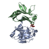
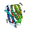
- PDB-2dwn: Crystal structure of the PriA protein complexed with oligonucleotides -
+ Open data

- Basic information
| Entry | Database: PDB / ID: 2dwn | |||||||||
|---|---|---|---|---|---|---|---|---|---|---|
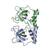
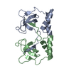
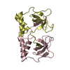
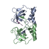
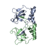
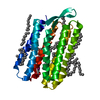
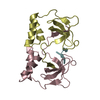
| Title | Crystal structure of the PriA protein complexed with oligonucleotides | |||||||||
 Components Components |
| |||||||||
 Keywords Keywords | HYDROLASE/DNA /  PROTEIN-DNA COMPLEX / HYDROLASE-DNA COMPLEX PROTEIN-DNA COMPLEX / HYDROLASE-DNA COMPLEX | |||||||||
| Function / homology |  Function and homology information Function and homology informationDnaB-DnaC-DnaT-PriA-PriC complex / DnaB-DnaC-DnaT-PriA-PriB complex / plasmid maintenance / primosome complex / DNA replication, synthesis of primer / 3'-5' DNA helicase activity / replication fork processing / DNA unwinding involved in DNA replication / DNA replication initiation / helicase activity ...DnaB-DnaC-DnaT-PriA-PriC complex / DnaB-DnaC-DnaT-PriA-PriB complex / plasmid maintenance / primosome complex / DNA replication, synthesis of primer / 3'-5' DNA helicase activity / replication fork processing / DNA unwinding involved in DNA replication / DNA replication initiation / helicase activity / response to gamma radiation / Hydrolases; Acting on acid anhydrides; Acting on acid anhydrides to facilitate cellular and subcellular movement / DNA-templated DNA replication / double-strand break repair / DNA recombination / DNA replication / hydrolase activity / response to antibiotic / DNA binding / zinc ion binding / ATP bindingSimilarity search - Function | |||||||||
| Biological species |  Escherichia coli (E. coli) Escherichia coli (E. coli)Synthetic construct (others) | |||||||||
| Method | X-RAY DIFFRACTION / SYNCHROTRON / MOLECULAR REPLACEMENT / Resolution: 3.35 Å | |||||||||
 Authors Authors | Sasaki, K. / Ose, T. / Tanaka, T. / Masai, H. / Maenaka, K. / Kohda, D. | |||||||||
 Citation Citation | Journal: EMBO J. / Year: 2007 Title: Structural basis of the 3'-end recognition of a leading strand in stalled replication forks by PriA. Authors: Sasaki, K. / Ose, T. / Okamoto, N. / Maenaka, K. / Tanaka, T. / Masai, H. / Saito, M. / Shirai, T. / Kohda, D. | |||||||||
| History |
|
- Structure visualization
| Structure viewer | Molecule: MolmilJmol/JSmol |
|---|
- Downloads & links
-Download
| PDBx/mmCIF format | 2dwn.cif.gz | 86.8 KB | Display | PDBx/mmCIF format |
|---|---|---|---|---|
| PDB format | pdb2dwn.ent.gz | 67.2 KB | Display | PDB format |
| PDBx/mmJSON format | 2dwn.json.gz | Tree view | PDBx/mmJSON format | |
| Others |  Other downloads Other downloads |
-Validation report
| Arichive directory | https://data.pdbj.org/pub/pdb/validation_reports/dw/2dwnftp://data.pdbj.org/pub/pdb/validation_reports/dw/2dwn | HTTPS FTP |
|---|
-Related structure data
| Related structure data |  2d7eSC  2d7gC 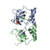 2d7hC  2dwlC  2dwmC S: Starting model for refinement C: citing same article ( |
|---|---|
| Similar structure data |

-Links
 PDBj
PDBj- Assembly
| Deposited unit | 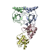
| ||||||||
|---|---|---|---|---|---|---|---|---|---|
| 1 | 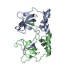
| ||||||||
| 2 | 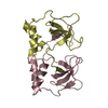
| ||||||||
| 3 | 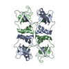
| ||||||||
| 4 | 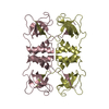
| ||||||||
| Unit cell |
|
-Components
| #1: Protein | Mass: 11776.790 Da / Num. of mol.: 4 / Fragment: Residues 1-105 Source method: isolated from a genetically manipulated source Source: (gene. exp.) Escherichia coli (strain K12) (bacteria)Strain: K12 / Gene: priA, b3935, JW3906 / Plasmid: pET15b / Species (production host): Escherichia coli / Production host: Escherichia coli BL21(DE3) (bacteria) / Strain (production host): BL21(DE3)References: UniProt: P17888, Hydrolases; Acting on acid anhydrides; Acting on acid anhydrides to facilitate cellular and subcellular movement#2: DNA chain | Mass: 597.455 Da / Num. of mol.: 2 / Source method: obtained synthetically / Source: (synth.) Synthetic construct (others) |
|---|
-Experimental details
-Experiment
| Experiment | Method: X-RAY DIFFRACTION / Number of used crystals: 1 |
|---|
- Sample preparation
| Crystal | Density Matthews: 3.3 Å3/Da / Density % sol: 62.77 % | ||||||||||||||||||||
|---|---|---|---|---|---|---|---|---|---|---|---|---|---|---|---|---|---|---|---|---|---|
| Crystal grow | Temperature: 293 K / Method: vapor diffusion, hanging drop / pH: 3.6 Details: 0.1M sodium citrate, 0.4M ammonium sulfate, pH 3.6, VAPOR DIFFUSION, HANGING DROP, temperature 293K | ||||||||||||||||||||
| Components of the solutions |
|
-Data collection
| Diffraction | Mean temperature: 100 K |
|---|---|
| Diffraction source | Source: SYNCHROTRON / Site: SPring-8  / Beamline: BL38B1 / Wavelength: 1 Å / Beamline: BL38B1 / Wavelength: 1 Å |
| Detector | Type: RIGAKU JUPITER 210 / Detector: CCD / Date: Apr 5, 2006 |
| Radiation | Protocol: SINGLE WAVELENGTH / Monochromatic (M) / Laue (L): M / Scattering type: x-ray |
| Radiation wavelength | Wavelength: 1 Å / Relative weight: 1 |
| Reflection | Resolution: 3.35→50 Å / Num. obs: 9392 / % possible obs: 99.9 % / Redundancy: 11 % / Biso Wilson estimate: 113.2 Å2 / Rsym value: 0.052 / Net I/σ(I): 15.4 |
| Reflection shell | Resolution: 3.35→3.47 Å / Redundancy: 11.3 % / Mean I/σ(I) obs: 2.73 / Num. unique all: 931 / Rsym value: 0.387 / % possible all: 100 |
- Processing
| Software |
| ||||||||||||||||||||
|---|---|---|---|---|---|---|---|---|---|---|---|---|---|---|---|---|---|---|---|---|---|
| Refinement | Method to determine structure: MOLECULAR REPLACEMENT Starting model: PDB ENTRY 2D7E Resolution: 3.35→20 Å / σ(F): 2.7
| ||||||||||||||||||||
| Refine analyze |
| ||||||||||||||||||||
| Refinement step | Cycle: LAST / Resolution: 3.35→20 Å
| ||||||||||||||||||||
| Refine LS restraints |
| ||||||||||||||||||||
| LS refinement shell | Resolution: 3.35→3.47 Å
|
