 Movie
Movie Controller

[English] 日本語
 Yorodumi
- PDB-6jo0: Crystal structure of the DTS-motif rhodopsin from Phaeocystis glo... -
+ Open data

- Basic information
| Entry | Database: PDB / ID: 6jo0 | ||||||
|---|---|---|---|---|---|---|---|
| Title | Crystal structure of the DTS-motif rhodopsin from Phaeocystis globosa virus 12T | ||||||
 Components Components | VirRDTS | ||||||
 Keywords Keywords |  MEMBRANE PROTEIN / microbial type rhodopsin from virus MEMBRANE PROTEIN / microbial type rhodopsin from virus | ||||||
| Function / homology |  Function and homology information Function and homology information | ||||||
| Biological species |  Phaeocystis globosa virus 12T Phaeocystis globosa virus 12T | ||||||
| Method | X-RAY DIFFRACTION / SYNCHROTRON / MOLECULAR REPLACEMENT / Resolution: 1.651 Å | ||||||
 Authors Authors | Hosaka, T. / Kimura-Someya, T. / Shirouzu, M. | ||||||
 Citation Citation | Journal: Proc.Natl.Acad.Sci.USA / Year: 2019 Title: A distinct lineage of giant viruses brings a rhodopsin photosystem to unicellular marine predators. Authors: Needham, D.M. / Yoshizawa, S. / Hosaka, T. / Poirier, C. / Choi, C.J. / Hehenberger, E. / Irwin, N.A.T. / Wilken, S. / Yung, C.M. / Bachy, C. / Kurihara, R. / Nakajima, Y. / Kojima, K. / ...Authors: Needham, D.M. / Yoshizawa, S. / Hosaka, T. / Poirier, C. / Choi, C.J. / Hehenberger, E. / Irwin, N.A.T. / Wilken, S. / Yung, C.M. / Bachy, C. / Kurihara, R. / Nakajima, Y. / Kojima, K. / Kimura-Someya, T. / Leonard, G. / Malmstrom, R.R. / Mende, D.R. / Olson, D.K. / Sudo, Y. / Sudek, S. / Richards, T.A. / DeLong, E.F. / Keeling, P.J. / Santoro, A.E. / Shirouzu, M. / Iwasaki, W. / Worden, A.Z. | ||||||
| History |
|
- Structure visualization
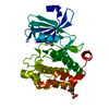
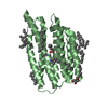
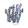
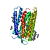
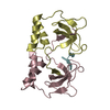
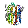
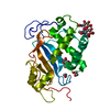
| Structure viewer | Molecule: MolmilJmol/JSmol |
|---|
- Downloads & links
-Download
| PDBx/mmCIF format | 6jo0.cif.gz | 120.1 KB | Display | PDBx/mmCIF format |
|---|---|---|---|---|
| PDB format | pdb6jo0.ent.gz | 90.7 KB | Display | PDB format |
| PDBx/mmJSON format | 6jo0.json.gz | Tree view | PDBx/mmJSON format | |
| Others |  Other downloads Other downloads |
-Validation report
| Arichive directory | https://data.pdbj.org/pub/pdb/validation_reports/jo/6jo0ftp://data.pdbj.org/pub/pdb/validation_reports/jo/6jo0 | HTTPS FTP |
|---|
-Related structure data
| Related structure data | 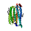 5b2nS S: Starting model for refinement |
|---|---|
| Similar structure data |


-Links
 PDBj
PDBj


- Assembly
| Deposited unit | 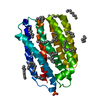
| ||||||||
|---|---|---|---|---|---|---|---|---|---|
| 1 |
| ||||||||
| Unit cell |
|
-Components
-Protein , 1 types, 1 molecules A
| #1: Protein | Mass: 28087.266 Da / Num. of mol.: 1 Source method: isolated from a genetically manipulated source Source: (gene. exp.) Phaeocystis globosa virus 12T / Gene: PGAG_00290 / Production host:  Escherichia coli (E. coli) / References: UniProt: G8DI69 Escherichia coli (E. coli) / References: UniProt: G8DI69 |
|---|
-Non-polymers , 9 types, 111 molecules 


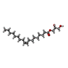

| #2: Chemical | ChemComp-RET / Retinal Mass: 284.436 Da / Num. of mol.: 1 / Source method: obtained synthetically / Formula: C20H28O Mass: 284.436 Da / Num. of mol.: 1 / Source method: obtained synthetically / Formula: C20H28O | ||||||||||||||
|---|---|---|---|---|---|---|---|---|---|---|---|---|---|---|---|
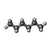
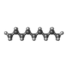
| #3: Chemical | ChemComp-OCT / Octane Mass: 114.229 Da / Num. of mol.: 6 / Source method: obtained synthetically / Formula: C8H18 Mass: 114.229 Da / Num. of mol.: 6 / Source method: obtained synthetically / Formula: C8H18#4: Chemical | ChemComp-PO4 / | Phosphate Mass: 94.971 Da / Num. of mol.: 1 / Source method: obtained synthetically / Formula: PO4 Mass: 94.971 Da / Num. of mol.: 1 / Source method: obtained synthetically / Formula: PO4#5: Chemical | ChemComp-HEX / Hexane Mass: 86.175 Da / Num. of mol.: 5 / Source method: obtained synthetically / Formula: C6H14 Mass: 86.175 Da / Num. of mol.: 5 / Source method: obtained synthetically / Formula: C6H14#6: Chemical | ChemComp-D10 / | Decane Mass: 142.282 Da / Num. of mol.: 1 / Source method: obtained synthetically / Formula: C10H22 Mass: 142.282 Da / Num. of mol.: 1 / Source method: obtained synthetically / Formula: C10H22#7: Chemical | ChemComp-D12 / | Dodecane Mass: 170.335 Da / Num. of mol.: 1 / Source method: obtained synthetically / Formula: C12H26 Mass: 170.335 Da / Num. of mol.: 1 / Source method: obtained synthetically / Formula: C12H26#8: Chemical | Nonane Mass: 128.255 Da / Num. of mol.: 3 / Source method: obtained synthetically / Formula: C9H20 Mass: 128.255 Da / Num. of mol.: 3 / Source method: obtained synthetically / Formula: C9H20#9: Chemical | ChemComp-OLB / ( |  Mass: 356.540 Da / Num. of mol.: 1 / Source method: obtained synthetically / Formula: C21H40O4 Mass: 356.540 Da / Num. of mol.: 1 / Source method: obtained synthetically / Formula: C21H40O4#10: Water | ChemComp-HOH / | WaterMass: 18.015 Da / Num. of mol.: 92 / Source method: isolated from a natural source / Formula: H2O |
-Experimental details
-Experiment
| Experiment | Method: X-RAY DIFFRACTION / Number of used crystals: 1 |
|---|
- Sample preparation
| Crystal | Density Matthews: 2.79 Å3/Da / Density % sol: 55.87 % |
|---|---|
| Crystal grow | Temperature: 293 K / Method: lipidic cubic phase / pH: 5.5 Details: Na-citrate (pH 5.5), 150 mM Na-phosphate, 25% PEG 600 |
-Data collection
| Diffraction | Mean temperature: 100 K / Serial crystal experiment: N | ||||||||||||||||||||||||
|---|---|---|---|---|---|---|---|---|---|---|---|---|---|---|---|---|---|---|---|---|---|---|---|---|---|
| Diffraction source | Source: SYNCHROTRON / Site: SPring-8  / Beamline: BL32XU / Wavelength: 1 Å / Beamline: BL32XU / Wavelength: 1 Å | ||||||||||||||||||||||||
| Detector | Type: DECTRIS EIGER X 9M / Detector: PIXEL / Date: Dec 8, 2016 | ||||||||||||||||||||||||
| Radiation | Protocol: SINGLE WAVELENGTH / Monochromatic (M) / Laue (L): M / Scattering type: x-ray | ||||||||||||||||||||||||
| Radiation wavelength | Wavelength: 1 Å / Relative weight: 1 | ||||||||||||||||||||||||
| Reflection | Resolution: 1.65→45.19 Å / Num. obs: 37658 / % possible obs: 99.9 % / Redundancy: 6.6 % / Biso Wilson estimate: 24.21 Å2 / CC1/2: 0.998 / Rmerge(I) obs: 0.086 / Rpim(I) all: 0.036 / Rrim(I) all: 0.094 / Net I/σ(I): 10.2 | ||||||||||||||||||||||||
| Reflection shell | Diffraction-ID: 1
|
- Processing
| Software |
| ||||||||||||||||||||||||||||||||||||||||||||||||||||||||||||||||||||||||||||||||||||||||||||||||||||||||||||||||||||||||||||||||||||||||||||||||||||||
|---|---|---|---|---|---|---|---|---|---|---|---|---|---|---|---|---|---|---|---|---|---|---|---|---|---|---|---|---|---|---|---|---|---|---|---|---|---|---|---|---|---|---|---|---|---|---|---|---|---|---|---|---|---|---|---|---|---|---|---|---|---|---|---|---|---|---|---|---|---|---|---|---|---|---|---|---|---|---|---|---|---|---|---|---|---|---|---|---|---|---|---|---|---|---|---|---|---|---|---|---|---|---|---|---|---|---|---|---|---|---|---|---|---|---|---|---|---|---|---|---|---|---|---|---|---|---|---|---|---|---|---|---|---|---|---|---|---|---|---|---|---|---|---|---|---|---|---|---|---|---|---|
| Refinement | Method to determine structure: MOLECULAR REPLACEMENT Starting model: 5B2N Resolution: 1.651→42.16 Å / SU ML: 0.19 / Cross valid method: THROUGHOUT / σ(F): 1.33 / Phase error: 21.83
| ||||||||||||||||||||||||||||||||||||||||||||||||||||||||||||||||||||||||||||||||||||||||||||||||||||||||||||||||||||||||||||||||||||||||||||||||||||||
| Solvent computation | Shrinkage radii: 0.9 Å / VDW probe radii: 1.11 Å | ||||||||||||||||||||||||||||||||||||||||||||||||||||||||||||||||||||||||||||||||||||||||||||||||||||||||||||||||||||||||||||||||||||||||||||||||||||||
| Displacement parameters | Biso max: 87.63 Å2 / Biso mean: 31.9584 Å2 / Biso min: 17.66 Å2 | ||||||||||||||||||||||||||||||||||||||||||||||||||||||||||||||||||||||||||||||||||||||||||||||||||||||||||||||||||||||||||||||||||||||||||||||||||||||
| Refinement step | Cycle: final / Resolution: 1.651→42.16 Å
| ||||||||||||||||||||||||||||||||||||||||||||||||||||||||||||||||||||||||||||||||||||||||||||||||||||||||||||||||||||||||||||||||||||||||||||||||||||||
| LS refinement shell | Refine-ID: X-RAY DIFFRACTION / Rfactor Rfree error: 0 / Total num. of bins used: 13
| ||||||||||||||||||||||||||||||||||||||||||||||||||||||||||||||||||||||||||||||||||||||||||||||||||||||||||||||||||||||||||||||||||||||||||||||||||||||
| Refinement TLS params. | Method: refined / Refine-ID: X-RAY DIFFRACTION
| ||||||||||||||||||||||||||||||||||||||||||||||||||||||||||||||||||||||||||||||||||||||||||||||||||||||||||||||||||||||||||||||||||||||||||||||||||||||
| Refinement TLS group |
|
