 Movie
Movie Controller

[English] 日本語
 Yorodumi
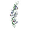
- PDB-5zvk: Crystal Structure of the Human Coronavirus MERS HR1 motif in comp... -
+ Open data

- Basic information
| Entry | Database: PDB / ID: 5zvk | ||||||
|---|---|---|---|---|---|---|---|
| Title | Crystal Structure of the Human Coronavirus MERS HR1 motif in complex with pan-CoVs inhibitor EK1 | ||||||
 Components Components |
| ||||||
 Keywords Keywords |  VIRAL PROTEIN/INHIBITOR / MERS / Spike protein / S2 domain / HR1 motif / pan-Coronavirus / VIRAL PROTEIN-INHIBITOR complex VIRAL PROTEIN/INHIBITOR / MERS / Spike protein / S2 domain / HR1 motif / pan-Coronavirus / VIRAL PROTEIN-INHIBITOR complex | ||||||
| Function / homology |  Function and homology information Function and homology informationendocytosis involved in viral entry into host cell / host cell endoplasmic reticulum-Golgi intermediate compartment membrane / membrane fusion / positive regulation of viral entry into host cell / receptor-mediated virion attachment to host cell / fusion of virus membrane with host plasma membrane / fusion of virus membrane with host endosome membrane / viral envelope / host cell plasma membrane / virion membrane / membraneSimilarity search - Function | ||||||
| Biological species |  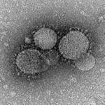 Middle East respiratory syndrome-related coronavirus Middle East respiratory syndrome-related coronavirussynthetic construct (others) | ||||||
| Method | X-RAY DIFFRACTION / SYNCHROTRON / MOLECULAR REPLACEMENT / Resolution: 3.31 Å | ||||||
 Authors Authors | Yan, L. / Yang, B. / Wilson, I.A. | ||||||
| Funding support |  China, 1items China, 1items
| ||||||
 Citation Citation | Journal: Sci Adv / Year: 2019 Title: A pan-coronavirus fusion inhibitor targeting the HR1 domain of human coronavirus spike. Authors: Xia, S. / Yan, L. / Xu, W. / Agrawal, A.S. / Algaissi, A. / Tseng, C.K. / Wang, Q. / Du, L. / Tan, W. / Wilson, I.A. / Jiang, S. / Yang, B. / Lu, L. | ||||||
| History |
|
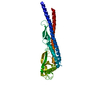
- Structure visualization
| Structure viewer | Molecule: MolmilJmol/JSmol |
|---|
- Downloads & links
-Download
| PDBx/mmCIF format | 5zvk.cif.gz | 130.7 KB | Display | PDBx/mmCIF format |
|---|---|---|---|---|
| PDB format | pdb5zvk.ent.gz | 104.9 KB | Display | PDB format |
| PDBx/mmJSON format | 5zvk.json.gz | Tree view | PDBx/mmJSON format | |
| Others |  Other downloads Other downloads |
-Validation report
| Arichive directory | https://data.pdbj.org/pub/pdb/validation_reports/zv/5zvkftp://data.pdbj.org/pub/pdb/validation_reports/zv/5zvk | HTTPS FTP |
|---|
-Related structure data
| Related structure data | 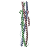 5zuvC 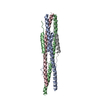 5zvmC  4modS C: citing same article ( S: Starting model for refinement |
|---|---|
| Similar structure data |


-Links
 PDBj
PDBj- Assembly
| Deposited unit | 
| ||||||||
|---|---|---|---|---|---|---|---|---|---|
| 1 |
| ||||||||
| Unit cell |
|
-Components
| #1: Protein | Mass: 8561.517 Da / Num. of mol.: 3 / Fragment: UNP residues 984-1062 Source method: isolated from a genetically manipulated source Source: (gene. exp.) Middle East respiratory syndrome-related coronavirusProduction host:  Escherichia coli (E. coli) / References: UniProt: W6A0A7, UniProt: K9N5Q8*PLUS Escherichia coli (E. coli) / References: UniProt: W6A0A7, UniProt: K9N5Q8*PLUS#2: Protein/peptide | Mass: 4923.550 Da / Num. of mol.: 3 Source method: isolated from a genetically manipulated source Details: The manually designed EK1 inhibitor was modified from a Coronavirus sequence (UNP P36334 SPIKE_CVHOC, UNP residues 1252-1288). Source: (gene. exp.) synthetic construct (others) / Production host: Escherichia coli (E. coli)Sequence details | The protein used for crystallization is a HR1-SGGRGG-EK1 fusion protein that contains MERS HR1 ...The protein used for crystallization is a HR1-SGGRGG-EK1 fusion protein that contains MERS HR1 motif, followed by SGGRGG linker, followed by manually designed EK1 peptide. | |
|---|
-Experimental details
-Experiment
| Experiment | Method: X-RAY DIFFRACTION / Number of used crystals: 1 |
|---|
- Sample preparation
| Crystal | Density Matthews: 2.27 Å3/Da / Density % sol: 45.86 % |
|---|---|
| Crystal grow | Temperature: 293 K / Method: evaporation / pH: 7 / Details: 0.2M KSCN, pH 7.0, 20% PEG3350 |
-Data collection
| Diffraction | Mean temperature: 100 K |
|---|---|
| Diffraction source | Source: SYNCHROTRON / Site: SSRF / Beamline: BL19U1 / Wavelength: 0.9785 Å |
| Detector | Type: DECTRIS PILATUS 6M / Detector: PIXEL / Date: Jan 3, 2017 |
| Radiation | Protocol: SINGLE WAVELENGTH / Monochromatic (M) / Laue (L): M / Scattering type: x-ray |
| Radiation wavelength | Wavelength: 0.9785 Å / Relative weight: 1 |
| Reflection | Resolution: 3.31→25.8 Å / Num. obs: 6009 / % possible obs: 99.9 % / Redundancy: 11.7 % / Biso Wilson estimate: 109 Å2 / CC1/2: 0.897 / Rpim(I) all: 0.036 / Rrim(I) all: 0.122 / Net I/σ(I): 20.7 |
| Reflection shell | Resolution: 3.31→3.43 Å / Mean I/σ(I) obs: 2.5 / Num. unique obs: 564 / Rpim(I) all: 0.324 / Rrim(I) all: 0.775 / % possible all: 100 |
- Processing
| Software |
| ||||||||||||||||||||||||||||||||||||||||
|---|---|---|---|---|---|---|---|---|---|---|---|---|---|---|---|---|---|---|---|---|---|---|---|---|---|---|---|---|---|---|---|---|---|---|---|---|---|---|---|---|---|
| Refinement | Method to determine structure: MOLECULAR REPLACEMENT Starting model: 4mod Resolution: 3.31→25.8 Å / SU ML: 0.44 / Cross valid method: FREE R-VALUE / σ(F): 1.38 / Phase error: 37.17
| ||||||||||||||||||||||||||||||||||||||||
| Solvent computation | Shrinkage radii: 0.9 Å / VDW probe radii: 1.11 Å | ||||||||||||||||||||||||||||||||||||||||
| Refinement step | Cycle: LAST / Resolution: 3.31→25.8 Å
| ||||||||||||||||||||||||||||||||||||||||
| Refine LS restraints |
| ||||||||||||||||||||||||||||||||||||||||
| LS refinement shell | Resolution: 3.31→3.43 Å / Rfactor Rfree: 0.318 / Rfactor Rwork: 0.34 | ||||||||||||||||||||||||||||||||||||||||
| Refinement TLS params. | Method: refined / Origin x: 36.3284 Å / Origin y: 34.8555 Å / Origin z: 188.4057 Å
| ||||||||||||||||||||||||||||||||||||||||
| Refinement TLS group | Selection details: all |
