 Movie
Movie Controller


+ Open data

- Basic information
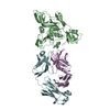
| Entry | Database: PDB / ID: 5yww | ||||||
|---|---|---|---|---|---|---|---|
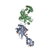
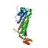
| Title | Archael RuvB-like Holiday junction helicase | ||||||
 Components Components | Nucleotide binding protein PINc | ||||||
 Keywords Keywords |  HYDROLASE / ATPase / Helicase HYDROLASE / ATPase / Helicase | ||||||
| Function / homology | Type II/IV secretion system protein / Type II/IV secretion system protein / Large family of predicted nucleotide-binding domains / PIN domain / P-loop containing nucleoside triphosphate hydrolase / Nucleotide binding protein PINc Function and homology information Function and homology information | ||||||
| Biological species |   Sulfolobus islandicus REY15A (acidophilic) Sulfolobus islandicus REY15A (acidophilic) | ||||||
| Method | X-RAY DIFFRACTION / SYNCHROTRON / MOLECULAR REPLACEMENT / Resolution: 2.33 Å | ||||||
 Authors Authors | Zhai, B. / Yuan, Z. / Han, X. / DuPrez, K. / Shen, Y. / Fan, L. | ||||||
 Citation Citation | Journal: Nucleic Acids Res. / Year: 2018 Title: The archaeal ATPase PINA interacts with the helicase Hjm via its carboxyl terminal KH domain remodeling and processing replication fork and Holliday junction. Authors: Zhai, B. / DuPrez, K. / Han, X. / Yuan, Z. / Ahmad, S. / Xu, C. / Gu, L. / Ni, J. / Fan, L. / Shen, Y. | ||||||
| History |
|
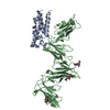
- Structure visualization
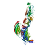
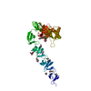
| Structure viewer | Molecule: MolmilJmol/JSmol |
|---|
- Downloads & links
-Download
| PDBx/mmCIF format | 5yww.cif.gz | 108.4 KB | Display | PDBx/mmCIF format |
|---|---|---|---|---|
| PDB format | pdb5yww.ent.gz | 80.7 KB | Display | PDB format |
| PDBx/mmJSON format | 5yww.json.gz | Tree view | PDBx/mmJSON format | |
| Others |  Other downloads Other downloads |
-Validation report
| Arichive directory | https://data.pdbj.org/pub/pdb/validation_reports/yw/5ywwftp://data.pdbj.org/pub/pdb/validation_reports/yw/5yww | HTTPS FTP |
|---|
-Related structure data
| Related structure data |  5f4hS S: Starting model for refinement |
|---|---|
| Similar structure data |

-Links
 PDBj
PDBj
- Assembly
| Deposited unit | 
| ||||||||
|---|---|---|---|---|---|---|---|---|---|
| 1 |
| ||||||||
| Unit cell |
|
-Components
| #1: Protein | Mass: 57153.012 Da / Num. of mol.: 1 / Mutation: R147K, I199S, R206A Source method: isolated from a genetically manipulated source Source: (gene. exp.) Sulfolobus islandicus REY15A (acidophilic)Strain: REY15A / Gene: SiRe_1432 / Production host:  Escherichia coli (E. coli) / References: UniProt: F0NID4 Escherichia coli (E. coli) / References: UniProt: F0NID4 |
|---|---|
| #2: Chemical | ChemComp-GOL / Glycerol  Mass: 92.094 Da / Num. of mol.: 1 / Source method: isolated from a natural source / Formula: C3H8O3 Mass: 92.094 Da / Num. of mol.: 1 / Source method: isolated from a natural source / Formula: C3H8O3 |
| #3: Water | ChemComp-HOH / Water Mass: 18.015 Da / Num. of mol.: 79 / Source method: isolated from a natural source / Formula: H2O Mass: 18.015 Da / Num. of mol.: 79 / Source method: isolated from a natural source / Formula: H2O |
-Experimental details
-Experiment
| Experiment | Method: X-RAY DIFFRACTION / Number of used crystals: 1 |
|---|
- Sample preparation
| Crystal | Density Matthews: 2.43 Å3/Da / Density % sol: 49.42 % |
|---|---|
| Crystal grow | Temperature: 287 K / Method: evaporation Details: 0.1M Bis-tris pH 6.5, 20% PEG monomethyl ether 5000 |
-Data collection
| Diffraction | Mean temperature: 100 K |
|---|---|
| Diffraction source | Source: SYNCHROTRON / Site: SSRF  / Beamline: BL17U1 / Wavelength: 0.979 Å / Beamline: BL17U1 / Wavelength: 0.979 Å |
| Detector | Type: ADSC QUANTUM 315r / Detector: CCD / Date: Jan 7, 2016 |
| Radiation | Protocol: SINGLE WAVELENGTH / Monochromatic (M) / Laue (L): M / Scattering type: x-ray |
| Radiation wavelength | Wavelength: 0.979 Å / Relative weight: 1 |
| Reflection | Resolution: 2.3→50 Å / Num. obs: 24438 / % possible obs: 99.4 % / Redundancy: 5.8 % / Rpim(I) all: 0.046 / Net I/σ(I): 32.38 |
| Reflection shell | Resolution: 2.33→2.39 Å / Redundancy: 5.7 % / Mean I/σ(I) obs: 2.44 / Rpim(I) all: 0.414 / % possible all: 97.1 |
- Processing
| Software |
| ||||||||||||||||||||||||||||||||||||||||||||||||||||||||||||||||||||||||||||||||||||||||||||||||||||||||||||||||||||||||||||||||||||||||||||||||||||||||||||||||||||||||||||||||||||||
|---|---|---|---|---|---|---|---|---|---|---|---|---|---|---|---|---|---|---|---|---|---|---|---|---|---|---|---|---|---|---|---|---|---|---|---|---|---|---|---|---|---|---|---|---|---|---|---|---|---|---|---|---|---|---|---|---|---|---|---|---|---|---|---|---|---|---|---|---|---|---|---|---|---|---|---|---|---|---|---|---|---|---|---|---|---|---|---|---|---|---|---|---|---|---|---|---|---|---|---|---|---|---|---|---|---|---|---|---|---|---|---|---|---|---|---|---|---|---|---|---|---|---|---|---|---|---|---|---|---|---|---|---|---|---|---|---|---|---|---|---|---|---|---|---|---|---|---|---|---|---|---|---|---|---|---|---|---|---|---|---|---|---|---|---|---|---|---|---|---|---|---|---|---|---|---|---|---|---|---|---|---|---|---|
| Refinement | Method to determine structure: MOLECULAR REPLACEMENT Starting model: 5F4H Resolution: 2.33→19.87 Å / Cor.coef. Fo:Fc: 0.948 / Cor.coef. Fo:Fc free: 0.94 / SU B: 8.336 / SU ML: 0.195 / Cross valid method: THROUGHOUT / ESU R: 0.404 / ESU R Free: 0.245 / Details: HYDROGENS HAVE BEEN ADDED IN THE RIDING POSITIONS
| ||||||||||||||||||||||||||||||||||||||||||||||||||||||||||||||||||||||||||||||||||||||||||||||||||||||||||||||||||||||||||||||||||||||||||||||||||||||||||||||||||||||||||||||||||||||
| Solvent computation | Ion probe radii: 0.8 Å / Shrinkage radii: 0.8 Å / VDW probe radii: 1.2 Å | ||||||||||||||||||||||||||||||||||||||||||||||||||||||||||||||||||||||||||||||||||||||||||||||||||||||||||||||||||||||||||||||||||||||||||||||||||||||||||||||||||||||||||||||||||||||
| Displacement parameters | Biso mean: 55.265 Å2
| ||||||||||||||||||||||||||||||||||||||||||||||||||||||||||||||||||||||||||||||||||||||||||||||||||||||||||||||||||||||||||||||||||||||||||||||||||||||||||||||||||||||||||||||||||||||
| Refinement step | Cycle: 1 / Resolution: 2.33→19.87 Å
| ||||||||||||||||||||||||||||||||||||||||||||||||||||||||||||||||||||||||||||||||||||||||||||||||||||||||||||||||||||||||||||||||||||||||||||||||||||||||||||||||||||||||||||||||||||||
| Refine LS restraints |
|